Skip to content

genetics

The MastAttack 107: The Layperson’s Guide to Understanding Mast Cell Diseases, Part 67

81. Is it true almost 20% of the population might have mast cell disease?

  • We need large scale studies in order to know for sure but this is very unlikely.
  • This figure first appeared in a paper published in 2013. This study asked mast cell patients, their relatives, and healthy control subjects to complete a questionnaire about whether or not they had symptoms of mast cell disease. 17% of the patients who were in the healthy control group reported symptoms that mast cell patients may experience. However, as we all know, you can have symptoms for a lot of reasons. Just sharing symptoms doesn’t mean anything. Also, surveys and questionnaires are known to be a bad way to collect hard data. By nature, they ask leading questions and are extremely subjective.
  • This same paper also tested for CKIT mutations in 20 mast cell patients and 20 healthy controls. The mast cell patient group included patients with MCAS, systemic mastocytosis, and cutaneous mastocytosis. The paper reported that 13/20 (65%) mast cell patients had mutations in the CKIT gene while 3/20 healthy controls (15%) had mutations in the CKIT gene. However, these mutations are not known to cause mast cell disease, or any diseases, for that matter. 20 people is a TINY number for a genetics study. You need a much larger group to really know whether or not a mutation occurs in healthy people or just people with a specific disease. We are talking hundreds to thousands of people needed in order to really know.
  • Many mastocytosis patients have a specific mutation called the CKIT D816V mutation. This mutation in mast cells only occurs when the patient has a clonal mast cell disorder like mastocytosis. People in the general population do not have the CKIT D816V without having mast cell disease.
  • Currently, the only CKIT mutations known to be associated with mast cell disease are the mutations at codon 816 of the CKIT gene. The D816V mutation is overwhelmingly the most common, but there are others that same exact spot, including D816Y, and a few others. Other mutations in the CKIT mutation are not known to be associated with mast cell disease.
  • Genes mostly tell your cells how to make proteins. The reason mutations can cause diseases is because they can change the structure of the protein that is made, affecting how it works. But not all mutations cause disease. Many mutations don’t change the proteins made by genes. This means that just having a mutation does not necessarily mean you have a disease. Mutations have to be linked to a disease by experimental work showing that people with a particular mutation have a particular disease.
  • Mutations are really very common. They occur so frequently that your cells have lots of failsafes in place specifically to work around or fix mutations. There are several ways they can do this but I’m just going to talk about one right now.
  • As I mentioned above, genes tell your cells how to make proteins. Genes are made of DNA. DNA is made of tiny building blocks called nucleotides. There are four kinds of DNA building blocks. These building blocks are grouped in bundles of three. Every one of those three DNA building block bundles tells the cell how to make a little piece of the protein. Those bundles are called codons. Then the three DNA building blocks next to that bundle tell the cell how to make another little piece of the protein, and so on. Those little pieces stick together and make the full sized protein.
  • The cell is able to make a protein by reading through this gene three pieces at a time. This is how our cells use genes to make proteins.
  • So now we know that those three DNA building blocks work together to me a tiny piece of protein. There are four kinds of DNA building blocks. What piece of protein is made is determined by which three building blocks are grouped together. There are many combinations of building blocks.
  • If one of those building blocks is mutated, it could cause the wrong protein piece to be made. So it seems that each of the three building blocks is really important to making the right protein. However, this is not the case. In fact, of those three building blocks, the last one is mostly irrelevant. If it’s mutated, it will usually still make the right protein piece. In some cases, the second one isn’t important either. The first one is the most important, but having a mutation there doesn’t always mean the protein is made wrong either.
  • So genes could potentially have up to 1/3 of its building blocks mutated without causing a problem. (I’m being very general here.) This phenomenon, in which the third DNA building block in a group can be wrong without messing up the protein is called wobble. Wobble is a built in mechanism that allows cells to make mutations sometimes without consequences.
  • That’s a lot of mutations that don’t cause problems. That’s a lot of mutations that still allow the cell to make the right protein. That’s a lot of mutations without causing symptoms or disease.
  • Your genes can withstand a lot of changes to single building blocks. When a single nucleotide is changed, it is called a single nucleotide polymorphism (snp). Most of these snps don’t cause trouble at all. The only way to know which ones cause problems is to gather up a bunch of people with these mutations and study them to see if they have diseases and, if so, which ones. So just having a mutation is not enough to know if it causes diseases without further study.

For additional reading, please visit the following posts:
Gene expression and the CKIT D816V mutation
The MastAttack 107: The Layperson’s Guide to Understanding Mast Cell Diseases, Part 2
The difference between C117+ and CKIT+
Heritable mutations in mastocytosis
The MastAttack 107: The Layperson’s Guide to Understanding Mast Cell Diseases, Part 2

I have answered the 107 questions I have been asked most in the last four years. No jargon. No terminology. Just answers.

3. What causes mast cell disease?

  • The cause of mast cell disease is not yet definitively known.
  • As mentioned yesterday, when the body makes too many copies of a broken cell, those cells are called ‘clonal’ cells. In clonal forms of mast cell disease, the bone marrow makes too many mast cells. Those mast cells also don’t work correctly. Examples of clonal mast cell diseases are systemic mastocytosis and cutaneous mastocytosis.
  • Patients with systemic mastocytosis often have a specific genetic mutation called the CKIT D816V mutation. About 80-90% of systemic mastocytosis patients have this mutation. This mutation is in mast cells and it tells the mast cells to stay alive WAY longer than they should. And mast cells already live for months or years, a very long time for cells to live in the body. So patients with this mutation can end up with way too many broken mast cells.
  • Despite the fact that we know that many patients have this mutation, we do not say that this mutation CAUSES the disease. The reason for this is that sometimes, mast cell patients don’t have the mutation when they get sick but they develop it later. Sometimes, mast cell patients have the mutation and then lose it later. So we are still looking for something that causes the disease.
  • Patients with non-clonal mast cell disease do not have a single major mutation like the CKIT D816V mutation. This makes it harder to diagnose. Researchers have found that many times, patients with MCAS DO have mutations similar to the ones systemic mastocytosis patients do. But the MCAS patients often have different mutations from each other. That’s why it’s not helpful yet for diagnosis.
  • Despite the fact that the mutations described here are not considered to be heritable, there is more and more evidence that mast cell disease can happen to many people in the same family. See the next question for more details.

4. Is mast cell disease heritable?

  • Mast cell disease often affects multiple members of the same family. Importantly, patients often have a different type of mast cell disease than their relatives. This implies that mast cell disease is more of a spectrum rather than several different diseases.
  • A survey found that 74% of mast cell patients interviewed reported at least one first degree relative that had mast cell disease. This same study found that 46% of those patients had mast cell disease that affected more than just their skin. This is called systemic disease.
  • The CKIT D816V mutation is the mutation most strongly associated with clonal mast cell disease. The CKIT D816V mutation is NOT heritable.
  • There are very rare instances of other heritable mutations in families that have mast cell disease. The significance of this is not clear.

5. Can mast cell disease be cured?

  • Generally speaking, there is no cure for mast cell disease.
  • Children who present with cutaneous mastocytosis sometimes grow out of their disease. Their lesions disappear. Their mast cell symptoms affecting the rest of the body may disappear. We do not know why this happens. It has been heavily researched with long term follow up of children with childhood mastocytosis (at least one paper followed them for 20 years).
  • Children with true systemic mastocytosis do not grow out of their disease.
  • There is not yet data on children with MCAS. Anecdotally, they do not seem to grow out of their disease like kids with cutaneous mastocytosis can. Importantly, this is just what it looks like to me. Again, there is no data.
  • People with adult onset mast cell disease have lifelong disease.
  • There is one notable exception to this scenario. There are reports of curing mast cell disease following hematopoietic stem cell transplant/bone marrow transplant.
  • Transplantation is EXTREMELY dangerous. The transplant is MUCH, MUCH more dangerous than mast cell disease. Many people do not survive the protocol necessary to prepare for transplant. Many die from complications, or from a disease they acquired after their transplant.
  • Rarely, people may have malignant forms of mast cell disease, aggressive systemic mastocytosis (ASM) or mast cell leukemia (MCL). A few patients with these diseases have tried transplants after everything else failed. While some did see improvement after transplant, no one has survived more than a few years.
  • Conversely, sometimes people with mast cell disease have these transplants for other reasons, like having another blood cancer or bone marrow disease that requires transplant. In this group of people, some see drastic improvement of their mast cell disease. Some see a full remission of mast cell disease. Some do not get any improvement. These findings are pretty recent so it’s hard to be more specific.

For more detailed reading, please visit these posts:

The Provider Primer Series: Introduction to Mast Cells

The Provider Primer Series: Mast cell activation syndrome (MCAS)

The Provider Primer Series: Cutaneous Mastocytosis/ Mastocytosis in the Skin

The Provider Primer Series: Diagnosis and natural history of systemic mastocytosis (ISM, SSM, ASM)

The Provider Primer Series: Diagnosis and natural history of systemic mastocytosis (SM-AHD, MCL, MCS)

Mast cell disease in families

Heritable mutations in mastocytosis

The Provider Primer Series: Mast cell activation syndrome (MCAS)

Mast cell activation syndrome (MCAS), also called mast cell activation disorder (MCAD), is an immunologic condition in which mast cells are aberrantly activated, resulting in inappropriate mediator release.

Presentation

  • MCAS can be responsible for chronic symptoms in multiple organs that cannot be attributed to another cause[vi].
  • Patients frequently receive diagnosis for a number of idiopathic conditions prior to correct diagnosis with MCAS[vi].
  • Mast cell activation syndrome is overwhelmingly a secondary condition. MCAS can be secondary to a number of conditions, including autoimmune diseases, connective tissue diseases, and atopic conditions[i].
  • The term “primary MCAS” refers to mediator release symptoms associated with mastocytosis[xvii] . However, the term “mastocytosis” generally conveys the understanding that both proliferation and mediator release symptoms are possible.
  • In idiopathic MCAS, no cause for symptoms can be identified[xvii] .
  • The presence of multiple mast cell patients in one family is not uncommon. A heritable gene has not yet been identified. Epigenetic mechanisms are suspected for transmission of mast cell disease to another generation[iv].
  • Approximately 75% of mast cell patients have at least one first degree relative with mast cell disease and not always the same subtype[ii]. For example, a mother may have MCAS, while one of her children has SM and the other has CM.

Diagnostic criteria

  • MCAS is a recently described diagnosis. In the absence of large studies, several groups have developed their own, sometimes conflicting, diagnostic criteria.
  • Differential diagnoses with potential to cause similar symptoms should be considered and excluded[iii].
  • The criteria most frequently used include those by a 2010 paper by Akin, Valent and Metcalfe[iii]; a 2011 paper by Molderings, Afrin and colleagues[iv]; and a 2013 paper by Castells and colleagues[v].
  • The criteria described in the 2011 paper by Molderings, Afrin and colleagues have been updated to include response to medication[vi].
  • Of note, a 2012 consensus proposal[x] was authored by a number of mast cell experts including Valent, Escribano, Castells, Akin and Metcalfe. It sees little practical use and is not generally accepted in the community.
  • The major sets of criteria listed above all include the following features:
    • Recurrent or chronic symptoms of mast cell activation
    • Objective evidence of excessive mast cell mediator release
    • Positive response to medications that inhibit action of mast cell mediators
  • Valent warns that in some cases, patients may not fulfill all criteria but still warrant treatment: “In many cases, only two or even one of these three criteria can be documented. In the case of typical symptoms, the provisional diagnosis of ‘possibly MCA/MCAS’ can be established, and in acute cases, immediate treatment should be introduced.”[vii]

Evidence of mediator release

  • Mast cells produce a multitude of mediators including tryptase, histamine, prostaglandin D2, leukotrienes C4, D4 and E4, heparin and chromogranin A[viii].
  • Serum tryptase and 24 hour urine testing for n-methylhistamine, prostaglandin D2, prostaglandin 9a,11b-F2 are frequently included in testing guidelines in literature (Castells 2013)[ix], (Akin 2010)[x], (Valent 2012)[xi].
  • It can be helpful to test for other mast cell mediators including 24 hour urine testing for leukotriene E4[xii]; plasma heparin[xiii]; and serum chromogranin A[xiv].
  • In most instances, elevation of a mediator must be present on two occasions[ix]. This helps to exclude situations of appropriate mast cell activation, such as infection or wound healing.
  • For patients with baseline tryptase level >15 ng/mL, elevation of tryptase above this baseline is only required on one occasion[viii].

Symptoms associated with mast cell activation

  • Mediator release causes a wide array of symptoms, including hypertension[xv], hypotension, hypertension, wheezing, itching, flushing, tachycardia, nausea, vomiting, diarrhea, constipation, headache, angioedema, fatigue, and neurologic symptoms[iv].
  • In a small MCAS cohort (18 patients), 17% had a history of anaphylaxis[xvii] . A larger data set is desirable.
  • Patients with history of anaphylaxis should be prescribed epinephrine autoinjectors[v]. If patient must be on a beta blocker, they should be prescribed a glucagon injector for use in the event of anaphylaxis[v].

Response to medications that inhibit action of mast cell mediators

  • Treatment of MCAS is complex and may require a number of medications. Second generation H1 antihistamines; H2 antihistamines; and mast cell stabilizers are mainstays of treatment[xvi].
  • Additional options include aspirin; anti-IgE; leukotriene blocker; and corticosteroids[xiii] .
  • First generation H1 antihistamines may be used for breakthrough symptoms[xiii] .
  • “An important point is that many different mediators may be involved in MCA-related symptoms so that the final conclusion the patient is not responding to antimediator therapy should only be drawn after having applied several different antimediator-type drugs[xiii] .
  • Inactive ingredients are often to blame for reaction to mast cell mediator focused medications. Many mast cell patients see benefit from having medications compounded[xvii].

Natural history

  • In one MCAS cohort of 18 patients, 33% had a complete (no unmanaged symptoms) response and 33% had a major (only one serious symptom) response after one year of mast cell treatment[xviii].
  • In another MCAS cohort of 135 patients, 51% demonstrated significant improvement, 11% had no obvious change in symptom severity and 38% experienced worsening symptoms[v]. (Author’s note: While described in an Afrin 2016[v] paper, the data from this cohort has not yet been published. Molderings is the principle investigator.


References

[i] Frieri M, et al. (2013). Mast cell activation syndrome: a review. Current Allergy and Asthma Reports, 13(1), 27-32.

[ii] Molderings GJ, et al. (2013). Familial occurrence of systemic mast cell activation disease. PLoS One, 8, e76241-24098785

[iii] Akin C, et al. (2010). Mast cell activation syndrome: proposed diagnostic criteria. J Allergy Clin Immunol, 126(6), 1099-1104.e4

[iv] Molderings GJ, et al. (2011). Mast cell activation disease: a concise practical guide for diagnostic workup and therapeutic options. Journal of Hematology & Oncology, 4(10), 10.1186/1756-8722-4-10

[v] Castells M, et al. (2013). Expanding spectrum of mast cell activation disorders: monoclonal and idiopathic mast cell activation syndromes. Clin Ther, 35(5), 548-562.

[vi] Afrin LB, et al. (2016). Often seen, rarely recognized: mast cell activation disease – a guide to diagnosis and therapeutic options. Annals of Medicine, 48(3).

[vii] Valent P. (2013). Mast cell activation syndromes: definition and classification. European Journal of Allergy and Clinical Immunology, 68(4), 417-424.

[viii] Theoharides TC, et al. (2012). Mast cells and inflammation. Biochimica et Biophysica Acta (BBA) – Molecular Basis of Disease, 1822(1), 21-33.

[ix] Picard M, et al. (2013). Expanding spectrum of mast cell activation disorders: monoclonal and idiopathic mast cell activation syndromes. Clinical Therapeutics, 35(5), 548-562.

[x] Akin C, et al. (2010). Mast cell activation syndrome: proposed diagnostic criteria. J Allergy Clin Immunol, 126(6), 1099-1104.e4

[xi] Valent P, et al. (2012). Definitions, criteria and global classification of mast cell disorders with special reference to mast cell activation syndromes: a consensus proposal. Int Arch Allergy Immunol, 157(3), 215-225.

[xii] Lueke AJ, et al. (2016). Analytical and clinical validation of an LC-MS/MS method for urine leukotriene E4: a marker of systemic mastocytosis. Clin Biochem, 49(13-14), 979-982.

[xiii] Vysniauskaite M, et al. (2015). Determination of plasma heparin level improves identification of systemic mast cell activation disease. PLoS One, 10(4), e0124912

[xiv] Zenker N, Afrin LB. (2015). Utilities of various mast cell mediators in diagnosing mast cell activation syndrome. Blood, 126(5174).

[xv] Shibao C, et al. (2005). Hyperadrenergic postural tachycardia syndrome in mast cell activation disorders. Hypertension, 45(3), 385-390.

[xvi] Cardet JC, et al. (2013). Immunology and clinical manifestations of non-clonal mast cell activation syndrome. Curr Allergy Asthma Rep, 13(1), 10-18.

[xvii] Afrin LB. “Presentation, diagnosis and management of mast cell activation syndrome.” In: Mast Cells. Edited by David B. Murray, Nova cience Publishers, Inc., 2013, 155-232.

[xviii] Hamilton MJ, et al. (2011). Mast cell activation syndrome: a newly recognized disorder with systemic clinical manifestations. Journal of Allergy and Clinical Immunology, 128(1), 147-152.e2

Mast cell mutations: SRSF2 in SM-AHNMD

SRSF2 is a splicing protein, which means it is involved in cutting RNA so that a gene makes the correct protein. SRSF2 mutations at position 95 (p95) have been found in 24-37% of SM patients. In nearly all cases, this mutation is found in patients that have SM-AHNMD. SRSF2 mutations at p95 are associated with myelodysplastic syndromes, leukemias and other hematologic diseases, including mastocytosis. After CKIT D816V mutation, it is the most common mutation found in systemic mastocytosis patients.

SRSF2 mutation occurs early in disease. One study indicates that it occurs in the time period between the initial TET2 mutation and the initial CKIT D816V mutation. Like TET2, it may predispose progenitor cells to develop subsequent mutations that cause disease. Patients with SRSF2 mutations are more likely to also have a TET2 mutation, and even more likely to have a mutation affecting another gene with epigenetic activity. SRSF2 alone does not drive neoplastic behavior in mast cells. SRSF2 has been found in patients without the CKIT mutation.

Importantly, SFSR2 is strongly associated with patients who develop SM-AHNMD. One study with a cohort of 72 patients with various forms of mastocytosis, including clonal MCAS (monoclonal mast cell activation syndrome) found that of 17 SM-AHNMD patients, 15 (88%) were positive for SFSR2 mutation. The associated conditions included AML, CMML, MDS, MSD (AREB), MPN, MPN/MDS and Waldenstrom’s macroglobunemia. SRSF2 mutations are not associated with any specific AHNMD and it does not predict survival time. This cohort included three patients who tested positive for the mutation but developed an AHNMD later in the study.

In SM-AHNMD patients, the SRSF2 mutations were found in both mast cells and monocytes in the bone marrow. This indicates that the mutation may cause transformation in both disease portions: SM and the AHNMD.


References:

Hanssens K., et al. SRSF2-P95 Hotspot Mutation is Highly Associated with Advanced Forms of Mastocytosis and Mutations in Epigenetic Regulator Genes. Haematologica 2013 [Epub ahead of print.]

Schwaab, J., Schnittger, S., Sotlar, K., Walz, C., Fabarius, A., Pfirrmann, M., et al., 2013.Comprehensive mutational profiling in advanced systemic mastocytosis. Blood122 (October (14)), 2460–2466.

Soucie, E., Brenet, F., Dubreuil, P. Molecular basis of mast cell disease. Molecular Immunology 63 (2015) 55-60.

Mast cell mutations: TET2 and mutation profiles of aggressive subtypes

TET2 (Tet methylcytosine dioxygenase 2) is found to be mutated in 20.8-29% of SM patients. Of note, dozens of mutations have been identified in this gene, including missense, nonsense, frameshift and deletion mutations. These mutations cause formation of a defective and less active TET2 enzyme. TET2 is located at chromosome 4q24 and mutations at this location are associated in both MPN and MDS conditions.

TET2 is involved in DNA methylation and demethylation, although the exact nature of this involvement is not clear. When a methyl group is added to a cytosine at a specific place in front of a gene, the gene is turned off and is not expressed. This is called “methylation.” TET2 adds a hydroxyl group to 5-methylcytosine, but it is not well understood if this turns the gene off. TET2 may also be involved in demethylating DNA, or removing those specific methyl groups. It has been shown to be involved with DNA demethylation during bone development.

One study looked at the mutational profiles of patients with various forms of SM, including ISM, SSM, SM-AHNMD, ASM and MCL, all of whom were positive for CKIT D816V mutation. 15/39 had a TET2 mutation. None of those patients had ISM or SSM. Of those with an aggressive form and a TET2 mutation, 67% had more than one TET2 mutation.

In this study, 24/27 patients with advanced SM (SM-AHNMD, ASM, MCL) had mutations beyond the D816V mutation. 5/5 SM-AHNMD patients and 19/22 ASM or MCL patients had multiple mutations (CKIT and something else.) In contrast, only 3/12 ISM or SSM patients had additional mutations. In advanced SM, 78% had at least 3 mutations, and 41% had at least 5.

These mutational profiles have clear implications clinically. 96% patients with major blood abnormalities (anemia <10 g/dL and/or thrombocytopenia < 100 x 10e9/L in addition to monocytosis > 1 x 10e9/L and/or eosinophilia >10%) had at least one additional molecular mutation regardless of SM subtype.

Advanced SM patients in this study all had one of the following multiple mutation profiles: 26% KIT-TET2-SRSF2, 18% KIT-SRSF2-RUNX1, 13% KIT-TET2-CBL, 10% KIT-SRSF2-ASXL1 10%, and 10% KIT-TET2-ASXL1. Patients with advanced SM (and therefore multiple mutations) were also found to be significantly older (68 years of age on average) than those with just the CKIT mutation (48 years of age on average.)

Having a TET2 mutation seems to predispose myeloid cells to become neoplastic later in life. It is important to distinguish that the TET2 mutation seems to “allow” this transformation rather than causing it. In mice who don’t have the TET2 gene and thus don’t have the TET2 enzyme, stem and progenitor cells have trouble maintaining balance and spontaneously become neoplastic later in life. In TET2 deficient cells, mast cells with D816V mutation show increase in proliferation and survival as opposed to those without with normal TET2 levels. Presence of TET2 in addition to the presence of CKIT D816V mutation is associated with more aggressive forms of SM (including ASM, MCL and SM-AHNMD.)


References:

Damaj, G., Joris, M., Chandersris, O., Hanssens, K., Soucie, E., Canioni, D., et al., 2014.ASXL1 but not TET2 Mutations Adversely Impact Overall Survival of PatientsSuffering Systemic Mastocytosis with Associated Clonal Hematologic Non-Mast-Cell Diseases. PLoS ONE 9 (1), e85362.

Schwaab, J., Schnittger, S., Sotlar, K., Walz, C., Fabarius, A., Pfirrmann, M., et al., 2013.Comprehensive mutational profiling in advanced systemic mastocytosis. Blood122 (October (14)), 2460–2466.

Soucie, E., Hanssens, K., Mercher, T., Georgin-Lavialle, S., Damaj, G., Livideanu, C.,et al., 2012. In aggressive forms of mastocytosis. TET2 loss cooperates with c-KITD816V to transform mast cells. Blood 120 (December (24)), 4846–4849.

Soucie, E., Brenet, F., Dubreuil, P. Molecular basis of mast cell disease. Molecular Immunology 63 (2015) 55-60.

Mast cell mutations: JAK2 and myeloproliferative neoplasms

Janus kinase 2 is also a tyrosine kinase like CKIT, but it is not a receptor on the outside of the cell. JAK2 is a helper protein that helps other molecules to send signals. It also affects the signaling of many clinically important molecules, like interleukin 3, interleukin 5, interleukin 5, interferon gamma, erythropoietin, thrombopoietin, growth hormone and prolactin. These molecules are involved in cell proliferation and the inflammatory response.

JAK2 mutation V617F seems to make hematopoietic cells more responsive to growth factors, causing excessive proliferation. JAK2 V617F is used as a marker for some Philadelphia negaitve myeloproliferative neoplasms (Ph- MPN), which include essential thrombocythemia (ET), an indolent disease in which too many platelets are produced; polycythemia vera (PV), in which too many red cells are produced; and myelofibrosis (MF), in which bone marrow is replaced with connective tissue that cannot make blood cells. JAK2 V617F is present in 40-50% of ET cases, 95% of PV cases, and 60% of MF cases.

In multiple studies, the frequency of JAK2 mutations in SM has ranged from 0-20% depending on the patient group. However, multiple studies have found a frequency of 4.2-5%, which is the generally accepted figure. JAK2 mutation is a strong predictor of myeloproliferative neoplasm but not necessarily of mastocytosis. However, where present in SM patients, it indicates a higher probability of developing another MPN.

In the control group of one study, 10 ISM patients without another myeloprofilerative neoplasm were negative for JAK2 mutation, and 15 MF patients without SM were negative for CKIT mutation. In the patient group, five people had both CKIT+ SM and MF. In four patients, the JAK2 V617F mutation was present. In the four patients with the JAK2 V617F mutation, it was found not only in the myelofibrosis cells (CD15+ myeloid cells), but also in the mast cells. In two of the patients, the CKIT mutation was found in the CD15+ myeloid cells of two patients. The data suggests that the JAK2 mutation may occur before the CKIT mutation in patients who have both SM and an associated hematologic disorder.

One study looked extensively at other mutations present in various types of CKIT+ systemic mastocytosis. The patient group was composed of 39 people, with 10 having ISM, 2 SSM, 5 SM-AHNMD, 15 ASM and 7 MCL. Only 2 patients out of the group were positive for JAK2 mutation. One patient had MCL, the other had SM-AHNMD. Both also had another myeloproliferative neoplasm (in the case of the SM-AHNMD patient, it was MDS.) This study found that presence of at least one other mutation in addition to CKIT D816V was associated with poorer prognosis, although presence of JAK2 V617F was not specifically identified as causing shorter lifespans.


References:

Schwaab, Juliana, et al. Comprehensive mutational profiling in advanced systemic mastocytosis. Blood 2013, 122 (14): 2460-2466.

Soucie, Erinn, et al. Molecular basis of mast cell disease. Molecular immunology 2015, 63 (1): 55-60.

J. Molderings. The genetic basis of mast cell activation disease – looking through a glass darkly. Critical Reviews in Oncology/Hematology 2014.

Sotlar, Karl, et al. Systemic mastocytosis associated with chronic idiopathic myelofibrosis. J Mol Diagn Jan 2008; 10(1): 58-66.

Heritable mutations in mastocytosis

While the most well-known mutation associated with SM is the CKIT D816V, there are numerous other mutations that can contribute to mast cell disease and presentation. The CKIT gene produces a tyrosine kinase receptor on the outside of the mast cell. Tyrosine kinases function as switches that turn certain cell functions on and off. When stem cell factor binds to the CKIT receptor, it turns on the signal for the mast cell to live longer than usual and to make more mast cells.

The D816V mutation is located in a specific part of the CKIT gene called exon 17. As many as 44% of SM patients have CKIT mutations outside of exon 17, either alone or in addition to the D816V mutation. (Please note that for the purposes of this post, SM is used to refer to SM, ASM and SM-AHNMD in keeping with the source literature.) Still, most doctors and researchers believe the D816V mutation is not heritable. This has important implications because it means many doctors also believe mast cell disease is sporadic and not heritable.

Almost 75% of MCAD (SM, MCAS, MCL) patients had at least one first degree relative with MCAD. This study, published in 2013, demonstrated that despite the non-heritable nature of the D816V mutation, mast cell disease is indeed heritable. Currently, four heritable mutations present in mast cell patients have been identified.

CKIT is often called KIT. In one family in which the mother, daughter and granddaughter have all have indolent SM, they were all found to have a deletion at position 409 in KIT (called KITdel409.) The KIT F522C mutation has been associated with ISM.   Another heritable mutation, KIT K509I, has been identified multiple times by different researchers. The first publication to identify this mutation was published in 2006. It has been found in a mother/daughter set who have ISM, and in another mother/daughter set in which the mother has ASM and the daughter has CM. This mutation was noted in a 2014 paper to be associated with well differentiated SM.

There have been reports of families in which multiple members with ISM or SM-AHNMD had the D816V mutation. Importantly, in these patients, the mutation was readily found in numerous cell types, including mast cells, CD34+ hematopoietic precursor cells, blood leukocytes, oral epithelial cells, blast cells and erythroid precursors. Despite this finding, the majority of literature continues to report the D816V mutation as not heritable.


References:

G.J. Molderings. The genetic basis of mast cell activation disease – looking through a glass darkly. Critical Reviews in Oncology/Hematology 2014.

G.J. Molderings, B. Haenisch, M. Bogdanow, R. Fimmers, M.M. Nöthen. Familial occurrence of systemic mast cell activation disease. PLoS One, 8 (2013), p. e76241

Hartmann, E. Wardelmann, Y. Ma, S. Merkelbach-Bruse, L.M. Preussenr, C. Woolery, et al. Novel germline mutation of KIT associated with familial gastrointestinal stromal tumors and mastocytosis. Gastroenterology, 129 (2005), pp. 1042–1046

R.A. Speight, A. Nicolle, S.J. Needham, M.W. Verrill, J. Bryon, S. Panter. Rare germline mutation of KIT with imatinib-resistant multiple GI stromal tumors and mastocytosis. J Clin Oncol, 31 (2013), pp. e245–e247

de Melo Campos, J.A. Machado-Neto, A.S.S. Duarte, R. Scopim-Ribeiro, F.F. de Carvalho Barra, J.Vassallo, et al.Familial mastocytosis: identification of KIT K509I mutation and its in vitro sensitivity to imatinib, dasatinib and PK412. Blood, 122 (2013), p. 5267

L.Y. Zhang, M.L. Smith, B. Schultheis, J. Fitzgibbon, T.A. Lister, J.V. Melo, et al. A novel K5091 mutation of KIT identified in familial mastocytosis – in vitro and in vivo responsiveness to imatinib therapy. Leukemia Res, 30 (2006), pp. 373–378

E.C. Chan, Y. Bai, A.S. Kirshenbaum, E.R. Fischer, O. Simakova, G. Bandara, et al. Mastocytosis associated with a rare germline KIT K509I mutation displays a well-differentiated mast cell phenotype. J Allergy Clin Immunol, 134 (2014), pp. 178–187

Akin, G. Fumo, A.S. Yavuz, P.E. Lipsky, L. Neckers, D.D. Metcalfe. A novel form of mastocytosis associated with a transmembrane c- Kit mutation and response to imatinib. Blood, 103 (2004), pp. 3222–3225

Escribano, R. Nunez-Lopez, M. Jara, A. Garcia-Montero, A. Prados, C. Teodosio, et al. Indolent systemic mastocytosis with germline D816 V somatic c-kit mutation evolving to an acute myeloid leukemia. J Allery Clin Immunol, 117 (Suppl.) (2006), p. S125

Food allergy series: Risk factors for developing food allergies

There are a number of factors that seem to contribute to developing food allergies. Genetics seems to play an important role. One study found that that 64% of monozygotic twins had a concordance of peanut allergy, while only 7% dizygotic twins concorded. This means that in 64% of identical twin sets, either both had peanut allergies or neither did, while that was the case in only 7% of fraternal twins. Because monozygotic twins have identical genetic sequences, this finding implies a strong genetic component. HLA haplotyping has been studied, with conflicting reports on links between HLA type and allergies.

One of the most important genetic findings regards filaggrin, a skin barrier protein. Patients with a specific filaggrin mutation are more likely to develop peanut sensitization. This indicates that a damaged skin barrier could cause food sensitization and allergy, and further supports the idea that non-oral exposures can be sensitizing. Additionally, filaggrin mutation causes increased inflammatory mediators in the skin.

Generally, children with peanut or tree nut allergies react the first time the food is ingested. It is thought that they previously encountered these allergens in their environment. Household exposure to peanut was a significant risk factor for peanut allergy in infants. Peanut responsive T cells are found in the skin homing T cells in peanut allergic patients, implying that patients may first be exposed through the skin. There is not yet enough data on maternal ingestion of allergens to know if this is a risk factor. There are conflicting data sets on whether breastfeeding is protective against food allergies, and in any case, outcome appears to be dependent on the mother’s own sensitivity profile. Now seen in multiple recent studies, it seems that early oral exposure to food allergens may actually be protective against food allergy, a change from data produced over a decade ago.

Immune dysregulation is obviously involved in food allergies. Low vitamin A and vitamin D, which modulate the immune system, have been noted as risk factors. Interestingly, food allergy frequency varies with latitude, indicating a further possible connection to sun exposure and vitamin D deficiency. High fat diet can also change the composition and behavior of the microbial content of the GI microbiome. Medium chain triglycerides can increase sensitization when given along with food antigens in mice. There are mixed results with long chain fatty acids.

The changes in hygiene, cleaning products and use of antimicrobial compounds by the general public in the last decades have been implicated in many of the immune changes we have seen, including increasing autoimmune diseases and food allergies. This is known as the hygiene hypothesis, and it states that reduced exposure to microbes changes immune defense, causing improper reactivity to harmless components, like food and self cells. In food allergic mice, the gut microbiota has a very specific composition and transferring this flora set can actually make others more likely to develop food allergies. Dysbiosis has been noted in children with food allergies and a sequencing study demonstrated that food allergic children with atopic dermatitis have reduced microbial diversity in the gut.


References:

Cecilia Berin, Hugh A. Sampson. Food allergy: an enigmatic epidemic. Trends in Immunology, Volume 34, Issue 8, August 2013, pages 390-397.

Cecilia Berin, Hugh A. Sampson. Mucosal Immunology of Food Allergy. Current Biology, Volume 23, Issue 9, May 2013, pages R389-R400.

MTHFR, folate metabolism and methylation

MTHFR (methylenetetrahydrofolate reductase) is an enzyme involved in folate metabolism. It is rate limiting, which means that if there is not enough, your body cannot metabolize enough folate; if there is too much, it metabolizes too much. How much folate your body is able to metabolize is directly related to how much MTHFR you have in your body.   Some folate broken down by your body is used to methylate DNA. There has been much conflicting evidence, but it seems that low folate in the body is associated with less DNA methylation overall, which may be associated with cancer (Crider, 2012.)

A single nucleotide polymorphism (SNP) is a change in DNA sequence in which one nucleotide (a DNA building block) is changed. Importantly, SNPs are common. In fact, they are so common that the way your body codes its DNA allows for SNPs to not change gene expression in many cases. This is called wobble. Three DNA building blocks in a row make one amino acid, which is used to build proteins that do things in the body. However, in many cases, the third building block can be any building block and it will still make the same amino acid. Please consider this. SNPs are so common that your cells know that in many cases, 1 in every 3 DNA building blocks can be anything and it won’t change a thing.

SNPs have become the topic of increased interest, both by medical and scientific professions and by lay people. It is certainly true that some SNPs play a role in development and progression of diseases. MTHFR has been found to have up to 24 reported SNPs, with two being of particular interest. These are C677T and A1298C.

The normal (or “wildtype”) form of MTHFR has a cytosine (C) nucleotide where people with the C677T mutation have a thymine nucleotide. If you have two copies of the regular gene, you are 677CC and homozygous for the regular form of the gene (the allele.) If you are 677CT, you have one copy of the allele with the SNP. If you are 677TT, you have two copies of the allele with the SNP. About 10% of North Americans are 677TT, meaning they have two copies of the mutated allele. It is most common in Hispanics and those of Mediterranean descent, next most common in Caucasians and least common in African Americans (Schneider, 1998.)

Being homozygous for 677TT can cause a mild MTHFR deficiency because that this form of the enzyme is generally less stable, which means it breaks down faster than usual. People with this profile are also more likely to develop mild hyperhomocysteinemia, an elevation of homocysteine in the blood. Homocysteine is consumed in the metabolism of folate, and because there is less MTHFR with the 677TT mutation, the homocysteine is not all getting used. It is also elevated in cases of B6, B12 and folic acid deficiency.

Increased homocysteine has been studied for its possible relationship to health issues, including increased clotting, strokes, schizophrenia and osteoporosis. Despite multiple studies (to be honest, quite a lot of studies), the results are really non-uniform. Because it was linked earlier to cardiovascular disease, multiple studies investigated the benefits of lowering homocysteine. In diabetic nephropathy patients, treating to lower homocysteine actually doubled cardiovascular events including heart attack and stroke, some leading to death, as well as decreased renal function (House, 2010.) But another study found lowering homocysteine decreased the risk of stroke by as much as 25% (Lonn, 2006.)

The effects of the C677T SNP have been most well studied in regards to development of cancers. Folic acid is a key metabolite in the development and proliferation of cells, so deficiency can be limiting to cell division. 677TT patients who are not folate deficient are actually 50% less likely to develop colorectal cancer or colorectal adenoma (Chen, 1999.) 677TT patients who are folate deficient have at least the same risk as 677CC patients and potentially more risk (Slattery, 1999.) 677TT patients are over four times less likely to develop acute lymphocytic leukemia. They have the same risk of developing acute myeloid leukemia (Skibola, 1999.) Some studies found them to have increased risk of cervical neoplasia, breast cancer, endometrial cancer and gastric cancer, but all of these studies were done on small populations (Xia, 2014.)

The other MTHFR SNP that gets a lot of attention is A1298C. At position 1298 of the MTHFR gene, the wildtype allele has adenine. In some people, it is substituted for cytosine. Wildtype is homozygous for adenine there and they are called 1298AA. If you have one copy of the SNP, you are 1298AC. If you have two copies, you are 1298CC. The A1298C has much less effect on the stability of the MTHFR protein than the C677T mutation. It is not known to cause elevation of homocysteine levels. There has been a lot of controversy over whether this mutation can cause a deficiency of BH4, tetrahydrobiopterin. BH4 is important in formation of neurotransmitters and nitric oxide, as well as consuming ammonia. Low levels of BH4 have been tied to phenylketonuria and trials with BH4 supplementation have seen encouraging results (Michals-Matalon, 2007.)

In the last six months or so, I have read about fifty scientific papers on MTHFR or related topics. I did this because I originally planned an MTHFR post for last summer. I didn’t do a post because the data is a mess. You cannot ascertain much of use from the peer reviewed literature on the C677T and A1298C mutations – and not for lack of effort. These mutations have been very well studied.

MTHFR mutations and methylation are talked about a lot in the mast cell community. Many people believe that having an MTHFR mutation severely impacts folate metabolism, which in turn means there is not enough methylation, and this dysregulation causes overexpression of genes causing disease. I have searched thoroughly for a link. Really thoroughly. I cannot find any link that is not the idea of one person and researched by that one person, usually outside of peer reviewed settings. I cannot find any link that is not described in detail by a person who does not stand to gain financially from patients who share their beliefs. I am not saying that MTHFR is definitely not linked to mast cell disease. I’m saying I can’t find any proof that it is. I can’t even find anything that SUGGESTS that it is. Might people with these mutations feel better with appropriate folic acid supplementation? Probably. Is that the same thing as causing mast cell disease? Certainly not. It is certainly not the same thing.

I think personal stories hold a lot of power. If your personal story is that your mast cell disease is significantly better controlled by addressing your MTHFR mutation, then I think that is fantastic. I think it is entirely possible that this is the case for many. But I do not believe it causes mast cell disease. And I think everyone would feel better with appropriate levels of folate, as it is an important player in many vital reactions.


References:

Schneider JA, Rees DC, Liu YT, Clegg JB (May 1998). “Worldwide distribution of a common methylenetetrahydrofolate reductase mutation”. Am. J. Hum. Genet. 62 (5): 1258–60

M L Slattery, et al. Methylenetetrahydrofolate reductase, diet and risk of colon cancer. Cancer Epidemiol Biomarkers Prev., 8 (1999), PP 560S-564S.

House, AA; Eliasziw, M; Cattran, DC; Churchill, DN; Oliver, MJ; Fine, A; Dresser, GK; Spence, JD (Apr 28, 2010). “Effect of B-vitamin therapy on progression of diabetic nephropathy: a randomized controlled trial.”. JAMA: the Journal of the American Medical Association 303 (16): 1603–9.

Lonn, E; Yusuf, S; Arnold, MJ; Sheridan, P; Pogue, J; Micks, M; McQueen, MJ; Probstfield, J; Fodor, G; Held, C; Genest J, Jr; Heart Outcomes Prevention Evaluation (HOPE) 2, Investigators (Apr 13, 2006). “Homocysteine lowering with folic acid and B vitamins in vascular disease.”. The New England Journal of Medicine 354 (15): 1567–77

Skibola CF, Smith MT, Kane E, Roman E, Rollinson S, Cartwright RA, Morgan G’ (October 1999). “Polymorphisms in the methylenetetrahydrofolate reductase gene are associated with susceptibility to acute leukemia in adults”. Proc. Natl. Acad. Sci. U.S.A. 96 (22): 12810–5.

Kim, Y I, et al. Methylenetetrahydrofolate reductase polymorphisms, folate and cancer risk: a paradigm of gene-nutrient interactions in carcinogenesis. Nutr Rev 58 (2000), pp 205-209.

Crider, Krista, et al. Folate and DNA Methylation: A Review of Molecular Mechanisms and the Evidence for Folate’s Role. Adv Nutr January 2012 Adv Nutr vol. 3: 21-38, 2012

Xia, Lei-Zhou, et al. Methylenetetrahydrofolate reductase C677T and A1298C polymorphisms and gastric cancer susceptibility. World J Gastroenterol. Aug 28, 2014; 20(32): 11429–11438.

J Chen, et al. MTHFR polymorphism, methyl-replete diets and the risk of colorectal carcinoma and adenoma among US men and women: an example of gene–environment interactions in colorectal tumorigenesis. J. Nutr., 129 (1999), pp. 560S–564S

DNA Methylation: How it works

DNA methylation is one of the ways your cells control which genes to express. It is an example of epigenetic modification. Epigenetics mechanisms like this do not change the DNA sequence, only the way the genes are expressed. Whether or not DNA methylation is heritable is not clear.

This is how DNA methylation works:

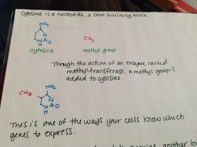
  • Cytosine is a nucleotide, a DNA building block.
  • Through the action of an enzyme called methyltransferase, a methyl group is added to cytosine.
  • This is one of the ways your cells know which genes to express.
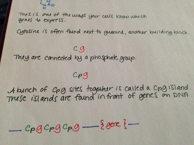
  • Cytosine is often found next to guanine, another building block. This is sometimes shown in literature as “CG.”
  • Cytosine and guanine are connected by a phosphate group. This is sometimes shown in literature as “CpG.”
  • A bunch of CpG sites together is called a CpG island. These islands are found in front of genes on DNA.
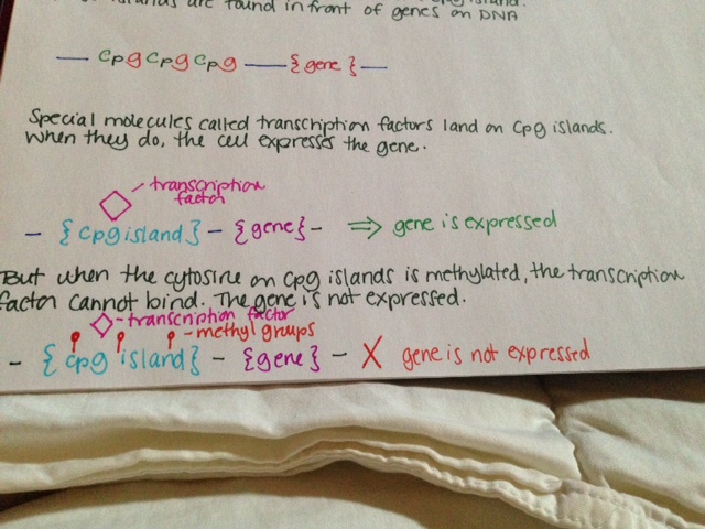
  • Special molecules called transcription factors land on CpG islands. When they do, the cell expresses the gene.
  • But when the cytosine on CpG islands is methylated, the transcription factor cannot bind. The gene is not expressed.

See pictures below.

Methylation is known to have an important role in cancer biology. Methylation of tumor suppressor genes causes the tumor suppressors not to be expressed, resulting in cancer.

Methylation 1Methylation 2Methylation 3