Skip to content

March 2015

Not a sad story

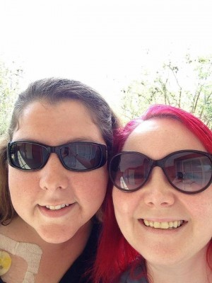
About a month ago, I had finally had enough of the oppressive snow wasteland known as Boston and I booked a trip to Florida. I told my masto friend I was coming down and we chirped excitedly about plans and what to do and all of those vacation things. It is no secret that my life has been generally frustrating recently so it seemed like this would provide an appropriate escape.

We made tentative plans for everything in the way only two people with mast cell disease can and generally derped out with excitement. We went back and forth about meds and supplies and safe foods. We decided to go to Disney.

Disney is one of my favorite places and a place I haven’t really had time to explore in several years. It also has an excellent reputation for accommodating health issues (including the very complex ones) and food allergies. We figured we would spend a couple days there, but I didn’t have high expectations for how much I would be able to do. Warm weather is a welcome change given the winter Boston just survived, but I don’t do well in heat, humidity or sunlight, especially when there is a lot of physical stress (like walking or standing for long periods of time). So I pretty much just hoped for the best in the same way I always do. A bad day at Disney is better than a bad day anywhere else.

I flew out of Boston on Thursday. It went pretty smoothly, with the exception of one woman who saw me get out of the wheelchair to walk into the bathroom. When I walked to the sink to wash my hands, she said, “You should be ashamed of yourself, there are people who really need those.” My reply was blistering and ended with, “People like you are the reason people like me kill themselves.” She was stunned to say the least.

Aside from this 90 seconds of unpleasantness, everything else was great. I touched down in Orlando around 6:15 on Thursday night. My friend Nikki picked me up and we checked into our room at the Port Orleans – French Quarter at Disney. It was so pretty. I know all Disney properties are pretty, but I liked this one a lot. We went out to a really nice, allergy safe dinner at a nearby hotel.

The next morning, we medded up and headed for Epcot. I LOVE Epcot. The only thing I wanted to do there was go to all the countries in the World Showcase. We got some (allergy safe) food and made it to all the countries before it started pouring. We covered our (accessed) ports and ran for the bus back to hotel.

It was a little hairy with gummy dressings and symptoms from the sudden temperature change, but we took meds and handled it. We took a nap back at the hotel and headed over to Magic Kingdom around 8. We went on the Haunted Mansion, It’s a Small World, Space Mountain and the new Seven Dwarfs Mine Ride, saw the fireworks and caught the end of the Electric Light Parade. We got back to the room around midnight and crashed hard, but in that exhausted way where you can’t sleep.

We had originally only booked two nights at Disney because we are crazy people who overestimated our physical capabilities. I figured that if we wanted to extend, it would be possible, even if we needed to switch hotels. I didn’t realize it was the start of Florida school vacation. We had called the Disney reservation line several times on Friday and they kept telling us nothing was available. Around 3am, I figured I would search online for available rooms since I wasn’t sleeping anyway and there was a room available at French Quarter! So I booked it and then we could not have to worry about waking up early to pack and also lack of sleep is one of my worst triggers so I had been worried about that. We got really lucky and slept in before heading to Animal Kingdom in the afternoon.

I have never seen a lot of the things at Animal Kingdom before so I was really excited. We went on the safari and saw lots of savannah animals, like giraffes, lions, zebras and hippos. We had booked fast passes for some rides so we did a lot of running around. (I should probably mention here that running is not something I do or tolerate well.) We went on this Mt. Everest roller coaster which was SO MUCH FUN (side note: at this point I learned that if you have a port and are on a roller coaster going backwards, it will feel like your port is pushing through your chest wall. It was really funny, when we started going backwards, both Nikki and I put our hands on our ports at the same time). We went back to the hotel and napped for a while and then took the ferry to Downtown Disney to get some food and watch Insurgent.

I was thoroughly fried by this point, and in that super uncomfortable, muscles hurt, about to react/actually reacting, nausea/vomiting space that I really hate. I was nauseous pretty much the whole time, but it was getting worse. I slept really late the next day and met Nikki at the MGM park, which I had also never seen in its entirety. We saw the Indiana Jones show, the Great Movie ride, a really cool stunt driving show and the Star Wars ride. At this point I was feeling the liquid courage effect of Benadryl so we waited in line (in the shade) for an hour for the Aerosmith Rockin’ Roller Coaster (which is one of my all time favorite roller coasters). It did not disappoint. Then there was only a short line for the Tower of Terror so we did that, too.

So, to summarize: two mast cell patients (who have had multiple surgeries, require regular IV meds and semi-regular epi, and have complicated food restrictions) went to Disney for three days, saw all four parks and Downtown Disney, ate food they didn’t prepare themselves and through the use of naps and liberal application of medication/IV fluids, were able to see/do literally all of the things they wanted to. LIKE FUCKING BOSSES.

Now we are at Nikki’s farm outside of Ocala which is very farmy and very beautiful. It is so calm here (except when the dogs and the pigs fight because the baby pig wants them to play with her, but still). I am recovering from the visceral adventures of Disney and feeling very glad that I came.

I try very hard to depict my life as realistically as possible, the good and the bad. I am in a place in my life right now where my life is hard a lot of the time and so that is what I write about. It’s not always my reality, it’s just my reality right now.

Everyone has hard things in their lives. I don’t think that being sick is any harder than losing a parent or a difficult divorce. It’s just different, and because my particular illness is unusual and uncommon, it seems worse to people. People say things to me sometimes, about how sad it makes them that I’m so young and so sick, or that I need a colostomy, or that I have a port, or whatever. They think my life is sad or tragic. My life is neither of those things.

I think sometimes that it’s easy to get stuck on how hard things are and how upsetting it is that you will never have your old life again. But we have these bright spots, and you can choose to elevate them in your mind so that they wash out the hard things, at least for a little while.

Don’t pity me, or people like me. This is my life, and it’s not a sad story.


Disney

Allergic effector unit: The interactions between mast cells and eosinophils

Eosinophils are granulocytes that can localize to the tissues under certain conditions, including allergic response. Eosinophilic granules contain the positively charged proteins major basic protein, eosinophil peroxidase, eosinophil cationic protein, and eosinophil-derived neurotoxin. Like mast cells, eosinophils release these granules in response to many things, including inflammatory signals, parasitic infection, tissue damage and allergic inflammation. They express many receptors, including receptors for platelet activating factor (PAF) and histamine receptors. PAF and histamine are both released by mast cells.

Mast cells and eosinophils are overwhelmingly found together in late and chronic stages of allergic inflammation. They function in such close concert that mast cells, eosinophils and their effects have been termed the allergic effector unit (AEU). Mast cells release signals that affect eosinophil behavior and receive signals from eosinophils. These cells often also function while in physical contact with one another. When eosinophils are in physical contact with mast cells, they live longer than normal. CD48, 2B4, DNAM-1 and Nectin-2 are all involved in the mast cell – eosinophil contact mechanism.

Major basic protein can activate mast cells and eosinophil peroxidase is taken up by mast cells as a signaling molecule. Tryptase draws eosinophils to mast cells and causes release of eosinophil peroxidase, IL-6 and IL-18 from eosinophils. Histamine and prostaglandin D2 also signal eosinophils to migrate towards mast cells. Mast cell secreted eotaxin activates eosinophils by the histamine 4 (H4) receptor. Both cell types secrete leukotrienes and both express leukotriene receptors.

When grown together, researchers are able to investigate the behavior of mast cells and eosinophils together. This is called co-culture. In 29% of cases, eosinophils will migrate towards resting (non-activated) mast cells. In 45% of cases, eosinophils will migrate towards IgE activated mast cells. In 47% of cases, eosinophils will migrate towards mast cells activated through a non-IgE pathway. The specific attractant signal has not been identified.

When co-cultured with eosinophils, basal mast cell mediator release was 5% higher. When the mast cells were activated by IgE, degranulation was 15% higher. In order to activate mast cells, eosinophils must be in contact with them. However, mast cells can activate eosinophils without contact. In co-cultures with mast cells, eosinophil peroxidase constituted 47% of eosinophil released proteins, compared with 18% normally.

In low term co-cultures, both mast cells and eosinophils stayed activated. TNF was high in the co-culture, but not IL-6, IL-8 and IL-10. Importantly, low relative numbers of mast cells could activate eosinophils, but mast cell activation was most effective when eosinophils were more numerous. Eosinophils are thought to reduce the threshold of mast cell responsiveness to IgE.


References:

Elishmereni M, Bachelet I, Nissim Ben Efraim AH, Mankuta D, Levi-Schaffer F. Interacting mast cells and eosinophils acquire an enhanced activation state in vitro. Allergy 2013; 68: 171–179.

Elishmereni M, Alenius HT, Bradding P, Mizrahi S, Shikotra A, Minai-Fleminger Y, et al. Physical interactions between mast cells and eosinophils: a novel mechanism enhancing eosinophil survival in vitro. Allergy 2011;66:376–385.

Minai-Fleminger Y, Elishmereni M, Vita F, Soranzo MR, Mankuta D, Zabucchi G et al. Ultrastructural evidence for human mast cell-eosinophil interactions in vitro. Cell Tissue Res 2010;341:405–415.

Puxeddu I, Ribatti D, Crivellato E, Levi- Schaffer F. Mast cells and eosinophils: a novel link between inflammation and angiogenesis in allergic diseases. J Allergy Clin Immunol 2005;116:531–536.

Mast cell mediators: Sphingosine-1-phosphate

Sphingosine-1-phosphate (S1P) is a lipid mediator involved in many processes, including development of vessels, vascular permeability, and immune function. It is found in the blood, often bound with proteins such as high density lipoprotein (HDL, “good cholesterol”). Receptors for S1P are found on many cell types.

Activation of the high affinity receptor for IgE causes production of S1P by mast cells. This may also affect the expression and activation of S1P receptors. Mast cells then secrete S1P into the surrounding space.  Mast cells also have receptors to bind S1P.

The S1P1 receptor helps to direct mast cells to sites of inflammation, but does not influence degranulation. The S1P2 receptor deters from localizing to sites of inflammation but enhances degranulation once they have migrated. S1P is known to increase during acute tissue inflammation, in airways following asthmatic challenge and in joints of rheumatic patients. S1P may be responsible for the accumulation of immune cells in such places, but the exact nature of this role is unclear.

S1P receptors regulate the vascular system, including heart rate and permeability.  S1P2 receptor makes vessels more permeable and regulates blood flow to various organs. S1P2 receptor is involved in counteracting the vasodilation effect of histamine (and thus low blood pressure). Histamine can stimulate S1P production.

S1P can also cause bradycardia and high blood pressure via the S1P3 receptor.  I am curious to know if S1P is involved in the high blood pressure type of anaphylaxis some people have.

In models where the genes for making S1P have been deleted, recovery from anaphylaxis is delayed, with severe hypotension. However, in mice with S1P2 receptors, injecting S1P could rescue mice from anaphylaxis. For this reason, molecules that can act on the S1P receptors are being investigated as possible drug targets to produce an alternative to epinephrine.


References:
Olivera A, Rivera J. An emerging role for the lipid mediator sphingosine-1-phosphate in mast cell effector function and allergic disease. Adv Exp Med Biol. 2011; 716: 123–142.

Allende ML, Proia RL. Sphingosine-1-phosphate receptors and the development of the vascular system. Biochim Biophys Acta. 2002;1582:222–227.

Olivera A, Eisner C, Kitamura Y, et al. Sphingosine kinase 1 and sphingosine-1-phosphate receptor 2 are vital to recovery from anaphylactic shock. J Clin Invest. 2010 “in press”.

Allergic to infections: Other behaviors of toll like receptors

I posted earlier this week about Toll-like receptors (TLRs). These are receptors on the outside of mast cells (and other cells) that tell them there is an infection. Instead of only being able to bind very specific molecules like receptors often do, these TLRs are able to bind lots of molecules that look alike. Once these are bound, it tells mast cells to activate, make mediators and release them.

After TLR2, TLR4 is the most well understood Toll-like receptor. Molecules that bind TLR4 are from infectious gram negative bacteria, several viruses (including RSV), Cryptococcus neoformans, and Candida albicans. It also binds fibrinogen, which is involved in the clotting cascade, and nickel. When infected with a gram negative bacteria, like E. coli or Ps. aeruginosa, mast cells secrete inflammatory molecules TNF, IL-6, IL-13, and IL-1b.

TLR4 also has a very intriguing behavior with opioid receptors on mast cells. These opioid receptors are the binding sites for opiate medications, like morphine, which are common triggers for mast cell patients. One study found that treatment with morphine actually interferes with TLR4 making inflammatory molecules. Other studies have found that opiates can bind TLR-4 directly. When bound, TLR-4 signals for release of TNFa, CCL1 and IL-5.

Other TLRs on mast cells can be bound by various molecules and produce and release mediators in return. TLR3 is bound by viral proteins and induces release of interferon a and b; TLR5 binds proteins from some flagellated bacteria and releases TNF and IL-1b; TLR9 binds unmethylated DNA of the type released by bacteria and DNA viruses, and releases interferon a, TNF, IL-1b and leukotrienes.

All TLR receptors can function independently of IgE. This is one example of an IgE independent pathway, or a way mast cells can degranulate or secrete mediators without IgE.


References:

Hilary Sandig and Silvia Bulfone-Paus. TLR signaling in mast cells: common and unique features. Front Immunol. 2012; 3: 185.

Abraham S. N, St John A. L. (2010). Mast cell-orchestrated immunity to pathogens. Nat. Rev. Immunol. 10440–452.

Dietrich N., Rohde M., Geffers R., Kroger A., Hauser H., Weiss S., Gekara N. O. (2010). Mast cells elicit proinflammatory but not type I interferon responses upon activation of TLRs by bacteria. Proc. Natl. Acad. Sci. U.S.A.1078748–8753

Gilfillan A. M., Tkaczyk C. (2006). Integrated signalling pathways for mast-cell activation. Nat. Rev. Immunol.6218–230.

Fehrenbach K., Port F., Grochowy G., Kalis C., Bessler W., Galanos C., Krystal G., Freudenberg M., Huber M. (2007). Stimulation of mast cells via FcvarepsilonR1 and TLR2: the type of ligand determines the outcome. Mol. Immunol. 442087–2094.

McCurdy,J.D., Olynych,T.J., Maher, L. H.,and Marshall, J.S.(2003). Cutting edge: distinct Toll-like receptor2 activators selectively induce different classes of mediator production from human mast cells. J. Immunol. 170, 1625–1629.

Medina-Tamayo, J., Ibarra-Sanchez, A., Padilla-Trejo,A., and Gonzalez- Espinosa, C. (2011). IgE-dependent sensitization increases responsiveness to LPS but does not modify development of endotoxin tolerance in mast cells. Inflamm. Res. 60, 19–27.

Qiao,H., Andrade,M.V., Lisboa,F. A., Morgan,K., and Beaven, M. A. (2006).FcepsilonR1 and toll-like receptors mediate synergistic signals to markedly augment production of inflammatory cytokines in murine mast cells. Blood 107, 610–618.

Yoshioka,M., Fukuishi,N., Iriguchi,S., Ohsaki, K., Yamanobe,H., Inukai, A., Kurihara,D., Imajo,N., Yasui, Y., Matsui, N., Tsujita, T., Ishii, A., Seya,T., Takahama,M., and Akagi, M. (2007). Lipoteichoicacid down- regulates FcepsilonRI expressionon human mast cells through Toll-like receptor2. J. Allergy Clin. Immunol. 120, 452–461.

Varadaradjalou, S., Feger, F., Thieblemont, N., Hamouda, N.B., Pleau, J. M., Dy,M., and Arock, M. (2003). Toll-likereceptor2 (TLR2)and TLR4 differentially activate human mast cells. Eur. J. Immunol. 33, 899–906.

My surgery with mast cell disease pro-tips

Surgery with mast cell disease is a bit of a logistical circus. I am fortunate to be able to have my surgeries at a major teaching hospital where my mast cell specialists are also located and mast cell disease is understood to some degree by most people. Even still, there are a lot of things that must be organized in order to ensure the safest procedure possible.

Surgery is an inherently triggering process. It’s stressful. Even if you know it’s coming and feel fine about it, it’s still stressful. It is mechanically triggering to the body due to manipulation of the skin and tissue damage and remodeling. Operating rooms (and hospitals generally) can be hard for mast cell patients, with chemicals everywhere, poor control over temperature and the use of things like blood pressure cuffs and masks. Please assume that surgery will trigger you and plan ahead. Do not expect to be able to undergo surgery without mast cell precautions.

The first thing to do is make sure all of your (relevant) doctors are on the same page. Schedule appointments with all of them. Do it face to face.

Schedule any testing with enough time to have conversations with all of these doctors. This is frustrating but necessary. You do not want to find out later that another option would have been better, or that there was a precaution that wasn’t taken.

Meet with the anesthesia team in person. If you are in the hospital/seen by hospital providers frequently, they will sometimes say that you don’t need a face to face pre-op because you have recent blood work/they already have your anesthesia protocol/whatever. Do not listen to them. When you meet with the anesthesia team, open with the fact that you have a rare, life threatening blood disorder that causes severe non-IgE mediated reactions. Say it like you mean it. Then ask them to go through their entire procedure with medications they will use. Tell them what medications cannot be used for you. Do not be shy. I bring a list of meds contraindicated in mast cell disease, as well as a list of meds that I personally react to. Give them this list.

If you have reason to believe that these doctors are not familiar with mast cell disease, bring them a review article.

After pre-op, I also prefer to meet with the surgeon again face to face to go over in excruciating detail what they will do and what materials will be used to do it. Give appropriate substitutions (betadine for chlorhexidine, or whatever). If you are unclear on this, please consult your mast cell specialist ahead of time. Ask them if the hospital stocks all of your meds and if they don’t, find out what alternative they use. Try these alternatives ahead of time whenever possible.

Discuss your dietary needs with your surgeon ahead of time.  Bring a list of foods and meals you can eat.  If they cannot make you food safely, you may have to have someone bring you food, but you do not want this to be a surprise.

If you need an interpreter, make sure this is very clear to everyone involved as early as possible.

Make sure your pain management plan is rock solid. If you need to trial pain meds in order to find a safe one before surgery, do it. Do NOT have surgery without a pain management plan. It delays healing and is very triggering for mast cell patients. Also, that just sucks.

Same for anti-nausea meds. You will get nauseous.

If your surgery requires prep of some kind, I strongly advise you to trial it before you need to use it. As an example, the typical bowel preps they use for colonoscopies and GI surgeries just do not work for me. You do not want to find that out two days before surgery.

If you are having bowel surgery, expect to react to the prep. It is physically stressful. Discussion adding medication during this period with your mast cell specialist. I take extra Benadryl and oral steroids during this period. If you are adrenally insufficient, please consult with your endocrinologist regarding the need for additional steroids.

Pick a date when your doctors do not have imminent out of town time coming up. (For me, I want my mast cell GI specialist, immunologist and surgeon to be available.) And make sure that whoever is covering their service during out of office hours understands mast cell disease! This is so important. Sometimes you have one doctor in a group who treats mast cell disease, you do not want to be talking to someone who specializes in food allergy when you are having trouble.

I often see that people have not followed the premedication protocol recommended for mast cell patients. Let me be very clear. You are going to react to surgery in some way. Premedication is not a suggestion. It is a requirement. This protocol is designed to suppress anaphylaxis and to allow your body to behave during the procedure and the 24 hours or so following. If you have a hard contraindication to one or part of the premedication protocol, ask your mast cell specialist what you should take instead. STEROIDS AND ANTIHISTAMINES SHOULD BE TAKEN AS PREMEDS FOR SURGERY 12 HOURS BEFORE THE PROCEDURE AND ONE HOUR BEFORE THE PROCEDURE. THIS IS NOT NEGOTIABLE.

The day of the surgery, bring your medications with you to the hospital in case they don’t have your brand/med/whatever. BRING YOUR EPIPENS.  If you use special dressings/have a central line/ostomy/G-tube/something, bring all your stuff for that.  They will probably not need it, but you never know.  Also bring a copy of your health care proxy/ advanced directive and your “greatest hits” (current meds, health conditions, allergies, etc). Try to get scheduled for the first surgery of the day, but remember that you will need to be there about two hours before they operate in order to get settled and get IV meds an hour before the procedure. Even if you schedule this ahead of time and no matter how many notes are written, someone is still going to give you a hard time about one or all of your premeds. Do not be dissuaded. These are compulsory.

If you have a bad feeling about what is happening, feel your necessary precautions are not being respected, or generally feel like you are being pushed into something you do not want to do, reschedule your surgery. I do not care if you are in your fancy hospital gown with your IV started. If you feel that you are not in the hands of providers who will take care of you, leave. Your safety is more important than convenience for these people. Your health is not a democracy.

If you are a mast cell patient, you are likely going to need some babysitting after surgery. I was not allowed to be alone for three weeks after my 2013 surgery. Schedule this ahead of time. Do not worry about entertaining people. Type up a list of your meds, what they do, how to take them, etc, for your babysitters.   Also type up a list of signs to watch for. MAKE SURE THAT ANYONE WHO IS TAKING CARE OF YOU KNOWS HOW TO USE AN EPIPEN.

Relax. Surgery is a bummer but with precautions, it is still very safe for mast cell patients.


Leptin: the obesity hormone released by mast cells

Leptin is a hormone that is primarily secreted by adipose tissue, but is also produced and released by mast cells. In turn, mast cells also have leptin receptors. Leptin is primarily known for its action of part of the hypothalamus to inhibit the hunger response. Importantly, the body responds forcefully to leptin levels by engaging both biological and behavioral mechanisms to conserve energy. It is seen by researchers as less of a “hunger satiety” signal and more of a “starvation” signal.

Patients with obesity often have higher circulating levels of leptin than those without obesity. This occurs because leptin is secreted by adipose tissue, which obese patients have in higher amounts due to their higher percentage of body fat. These people seem to be resistant to the chemical action of leptin, possibly through a change in activity of leptin receptors in the hypothalamus. Some studies suggest that in obese patients, less leptin leaves the blood stream and crosses into the brain.

Leptin is now known to have a variety of other effects on the body, including modulating the immune system. It activates inflammatory cells, promotes T cell responses and mediates production of TNF, IL-2 and IL-6. In many inflammation models, cells express more leptin receptors than usual. In diet induced obese mice, mast cells have been observed to store and secrete TNF. In immune mediated diseases like autoimmune diseases, circulating levels of leptin are increased, and this in turn translates to higher levels of inflammatory cytokines.

Interestingly, leptin suppresses signals from the IgE receptor to make mediators. In leptin receptor deficiency models, magnified IgE anaphylaxis was observed. Leptin also seems to control the number of mast cells through some unclear mechanism. In leptin deficient mice, mast cell density is significantly higher in abdominal lymph nodes and fat deposits.

Leptin influences the release of many other molecules, including ghrelin. Ghrelin is the “hunger hormone,” released in the stomach and possibly elsewhere. It stimulates the hunger response in the body and also acts on the hypothalamus. The relationship between leptin and ghrelin is very complex and still being elucidated. However, it is thought that high levels of circulating leptin suppress secretion of ghrelin. This is especially of interest in inflammatory conditions as ghrelin suppresses production of a number of inflammatory markers, including TNF, IL-8, MCP-1, IL-1b, IL-6, CRP and others. This effect is so pronounced that it is being investigated as a treatment option for many conditions. Ghrelin has also been observed in one study in induce mast cell activation through a receptor independent pathway.


References:

Baatar D, Patel K, Taub DD. The effects of ghrelin on inflammation and the immune system. Mol Cell Endocrinol. 2011 Jun 20; 340(1): 44-58.

Hirayama T, et al. Ghrelin and obestatin promote the allergic action in rat peritoneal mast cells as basic secretagogues. Peptides. 2010 Nov;31(11):2109-13

Klok MD, Jakobsdottir S, Drent ML. The role of leptin and ghrelin in the regulation of food intake and body weight in humans: a review. Obes Rev. 2007 Jan; 8(1): 21-34.

Taildeman J, et al. Human mast cells express leptin and leptin receptors. Histochem Cell Biol. 2009 Jun; 131(6): 703-11.

Patricia Fernández-Riejos, Souad Najib, Jose Santos-Alvarez, Consuelo Martín-Romero, Antonio Pérez-Pérez, Carmen González-Yanes, and Víctor Sánchez-Margalet. Role of Leptin in the Activation of Immune Cells. Mediators of Inflammation, Volume 2010 (2010), Article ID 568343, 8 pages.

Altintas et al. Leptin deficiency-induced obesity affects the density of mast cells in abdominal fat depots and lymph nodes in mice. Lipids in Health and Disease 2012, 11:21

Horizon

The last couple of months have been really stressful. Several of my work projects are all requiring a lot of attention right now. I am trying to iron out some details around MastAttack and make plans for the future. I live in the grey bleakness of New England in the midst of a record breaking winter. I am having some setbacks regarding GI function and pain.

And of course, I am having surgery soon. The amount of feelings I have about this surgery is surprising given the fact that I have always expected to have it. I don’t know. I guess it just seemed further off. The horizon seems so far away until you’re standing on the edge of the world, about to fall off.

Bowel surgery when you have mast cell disease is a complicated affair. I have to get buy in from all the relevant specialists and they all have to agree on a plan. I have to schedule surgery when everyone is in town and not taking vacation in the near future. I have to arrange care (nursing and otherwise) for weeks after I leave the hospital. I have to finish up several work things before they operate. I want to get some things lined up for MastAttack before I go.

My surgery was scheduled for April 28. I saw my surgeon this week to go over everything. He is not convinced that removing all of my colon is the best move. I am going to repeat some motility testing. Specialized testing generally takes weeks to get scheduled. Which would literally give us the results days before my scheduled surgery date, and that’s cutting it a little close for me.

I scheduled all my testing, then called my surgeon’s secretary. I rescheduled my surgery for mid-May. I am frustrated that there is still disagreement so close to my surgery date, but I understand why. We can’t just look and see what happened to the last twenty people like me who had their colons removed. There just isn’t anyone like me.

Part of why this whole production has been stressful is because I saw this coming a mile away. Needing my colon removed is not a surprise. We discussed removing more of my colon when I had my surgery in 2013 (I still have about 70% of my colon).   We weighed the pros and cons then, so I feel like having a similar conversation two years later shouldn’t generate so many questions. But things change, and my body has changed, and I have changed, as a person. What I want now is not the same as two years ago.

I thought pushing back the surgery would make me mad, but it really didn’t. It was a relief. I immediately felt calmer. It gives me time to make decisions without pressure. It gives me time to take some time for myself and focus on the things I care about.

If the amount of messages I have received are any indication, the weight of my life in recent months has been apparent to my readers. You guys are fantastic. You are so sweet and kind and respectful of my time and my feelings. I really feel so privileged to be part of this thing we are all doing together.

It has not escaped notice that MastAttack is turning into a much larger undertaking than I could ever have anticipated. I think some people are worried that I write these posts and answer questions out of a sense of obligation. A few weeks ago, I took a week off from the blog. I didn’t research or write anything about mast cell disease.

But at the end of the week, all I wanted to do was write posts about mast cells and diabetes. Not because I felt like people would be upset if I didn’t. Because I wanted to write it in case it was helpful. And because I like doing this.

In the last few months, this amazing thing has been happening. I am getting questions from people that are very nuanced, that show a really good understanding of the biology involved. Some of these questions are coming from people who have progressed a long way in their understanding of this disease. They are questioning me and bringing up findings from papers I haven’t read or correcting me when I get sloppy with the details. It is so cool. We are getting somewhere. This is getting to be bigger than me. And that was always the point.

I’m going to tell you guys a secret. I don’t want to be doing this in ten years. I don’t want to be writing articles or posts or reading literature constantly. In ten years, I want all this information to be available in a concise, easy to digest form for anyone to use. I want so many people to know everything that I know that I become obsolete. It’s starting, and you’re all part of that.

Along those lines, it’s time for me to get some help with all of this. I’m taking some people up on offers to help out, and will be asking for help with specific tasks in the upcoming months. If you think you might want to help, feel free to message me on FB or send me an email. There will be more details in future posts.

As for me, I’m feeling decidedly less stressed than I have in a while. All of my work stuff will either get done or it won’t, and all of my blog stuff will either get done or it won’t.   I’ll have surgery and I think it will help. And if it doesn’t, it doesn’t. The risk of ending up with a permanent ileostomy is scary, but not trying to remove a huge source of inflammation and live without an ostomy is scarier. You can’t be afraid to try.

In the meantime, I’m taking some time to relax before this next stage of my life begins. I’m going to Florida to visit a dear friend (and Disney!) later this week. The week I was supposed to have surgery, I will be going to California to visit Team Addie, my mastsister Addison and her family. And deciding to do those things felt warm and peaceful. It is exactly the right medicine for this weariness.

So I guess what I’m trying to say is that I appreciate all of your support. And I appreciate all of you individually more than you know. Every time I see someone jump in with the right answer or a message of support, I am honored to be part of this. And I’m so thankful and touched by your messages of concern and support. It’s nice to have people to catch you once in a while.

Sometimes things are hard, but everything’s gonna be okay. Okay, or better. It’s like my guarantee.


Allergic to infections: How bacteria, viruses and fungi activate mast cells

I am often asked about whether an infection, even a mild cold, can cause worsening mast cell symptoms.  The answer is yes.  Viral, fungal and bacterial infections can all cause mast cell activation, and patients with prior activated mast cells are especially susceptible.  This is why it is so important for mast cell patients to avoid contagious illness as much as possible.

Several cell types in the human body have Toll-like receptors (TLRs) on their cell surfaces. These receptors bind many types of molecules that indicate presence of infection. These molecules are called pathogen-associated molecular patterns (PAMPs) and they share similar shapes that identify them as being released by infecting organisms. When these PAMPs are bound by TLRs on cell surfaces, it sends signals for the cells to mount an immune response.

The expression of TLRs on mast cells has been well studied using both mouse (murine) and human mast cells. TLR1, 2, 3, 4, 5, 6, 7, 9 and 10 have been identified on mast cells by at least one study. Some of these TLRs were only detected by finding related mRNA. (When cells express a gene to make a protein like a TLR, the DNA gene is copied into mRNA, which tells the cell how to make the TLR.) Since only the mRNA and not the TLR was directly identified, these TLRs require more research to be fully characterized.

TLR2 is one of the most well studied and understood of toll-like receptors found on mast cell surfaces. TLR2 is also known as CD282. Substances that bind to TLR2 include many molecules released by bacteria and fungi. Several types of peptidoglycans and found in bacterial cell membranes bind TLR2. In particular, lipoteichoic acid is a potent activator of TLR2. This molecule is found on the surfaces of gram-positive bacteria, like Staphylococcus spp. (Staph, MRSA) and Streptococcus spp. (Strep). Other bacteria that are known to activate TLR2 include Neisseria meningitides, Haemophilus influenzae, and Borrelia burgdorferi, among others. Mycobacteria are also activating to TLR2. Zymosan is found in cell membranes of yeast and binds TLR2. Aspergillus fumigatus (fungi) and several viruses, including Herpes simplex, Varicella zoster, Cytomegalovirus and measles, activate TLR2 responses. Heat shock protein 70 (HSP 70) is released by cells in the body when they are under certain types of stress, and this can activate TLR2.

When TLR2 is bound, mast cells produce and release several types of molecules that are not prestored in granules. The molecules released depend on which protein has bound TLR2. These molecules include IL-1b, which causes inflammatory pain hypersensitivity; IL-5, which activates eosinophils; leukotriene B4, which forms reactive oxygen species and participates in inflammation; leukotriene C4, which causes slow contraction of smooth muscle, including in the airway; GM-CSF (Granulocyte macrophage colony-stimulating factor), a growth factor for white blood cells; TNF, which has many inflammatory effects; RANTES, which brings other white cells to the site of inflammation; and others. TLR2 activation is responsible for the worsening of asthma symptoms in the presence of bacterial infection.

Multiple studies reported that stimulation of TLR2 with peptidoglycan (a constituent of gram positive bacterial cell membranes) induced degranulation. Stimulation with peptidoglycan induced histamine release as well as cytokine release in a 2003 study (Varadaradjalou 2003). Another study found that peptidoglycan did not cause statistically significant degranulation, but zymosan (a fungal product) and Pam3Cys (a synthetic molecule that acts like LPS, another component of bacterial membranes) did induce significant degranulation (McCurdy 2003). Other studies have not been able to replicate these results.

There is also evidence that stimulation of TLR2 can change the behavior of mast cells. When mast cells are grown in the presence of bacterial cell membrane products, they make different amounts of different proteins. Another study demonstrated that two bacterial cell membrane products downregulated the amount of FceRI (the IgE receptor) on the surface of mast cells, so after two days, mast cells were less responsive to stimulation by IgE molecules. This was partially due to the effects of TLR2 (Yoshioka 2007).

However, mast cells that are sensitized react more strongly to TLR2 activation with LPS (Medina-Tamayo 2011). This effect seems to be reliant on prior binding of IgE. Other very technical studies have investigated the effect of antigen (such as bacterial, viral or fungal products) on the interplay between the IgE receptor and TLR receptors.   While most of this work has been done in mouse cells, several investigators have shown that activation of TLR receptors and the IgE receptor causes enhanced release of cytokines but not degranulation. It is thought that the exaggerated response to IgE receptor and TLR2 stimulation can cause the exacerbation of allergic type conditions during active infection. (Qiao 2006)


References:

Hilary Sandig and Silvia Bulfone-Paus. TLR signaling in mast cells: common and unique features. Front Immunol. 2012; 3: 185.

Abraham S. N, St John A. L. (2010). Mast cell-orchestrated immunity to pathogens. Nat. Rev. Immunol. 10440–452.

Dietrich N., Rohde M., Geffers R., Kroger A., Hauser H., Weiss S., Gekara N. O. (2010). Mast cells elicit proinflammatory but not type I interferon responses upon activation of TLRs by bacteria. Proc. Natl. Acad. Sci. U.S.A.1078748–8753

Gilfillan A. M., Tkaczyk C. (2006). Integrated signalling pathways for mast-cell activation. Nat. Rev. Immunol.6218–230.

Fehrenbach K., Port F., Grochowy G., Kalis C., Bessler W., Galanos C., Krystal G., Freudenberg M., Huber M. (2007). Stimulation of mast cells via FcvarepsilonR1 and TLR2: the type of ligand determines the outcome. Mol. Immunol. 442087–2094.

McCurdy,J.D., Olynych,T.J., Maher, L. H.,and Marshall, J.S.(2003). Cutting edge: distinct Toll-like receptor2 activators selectively induce different classes of mediator production from human mast cells. J. Immunol. 170, 1625–1629.

Medina-Tamayo, J., Ibarra-Sanchez, A., Padilla-Trejo,A., and Gonzalez- Espinosa, C. (2011). IgE-dependent sensitization increases responsiveness to LPS but does not modify development of endotoxin tolerance in mast cells. Inflamm. Res. 60, 19–27.

Qiao,H., Andrade,M.V., Lisboa,F. A., Morgan,K., and Beaven, M. A. (2006).FcepsilonR1 and toll-like receptors mediate synergistic signals to markedly augment production of inflammatory cytokines in murine mast cells. Blood 107, 610–618.

Yoshioka,M., Fukuishi,N., Iriguchi,S., Ohsaki, K., Yamanobe,H., Inukai, A., Kurihara,D., Imajo,N., Yasui, Y., Matsui, N., Tsujita, T., Ishii, A., Seya,T., Takahama,M., and Akagi, M. (2007). Lipoteichoicacid down- regulates FcepsilonRI expressionon human mast cells through Toll-like receptor2. J. Allergy Clin. Immunol. 120, 452–461.

Varadaradjalou, S., Feger, F., Thieblemont, N., Hamouda, N.B., Pleau, J. M., Dy,M., and Arock, M. (2003). Toll-likereceptor2 (TLR2)and TLR4 differentially activate human mast cells. Eur. J. Immunol. 33, 899–906.

Diabetes, steroids and hypoglycemia

Following alloxan induction of diabetes, rats overexpress glucocorticoids. This in turn depletes the mast cell populations in the skin, lungs and intestines. Glucocorticoids interfere with production and expression of tissue cytokines and stem cell factor, a growth factor for mast cells.

Several experiments have definitively proven that these steroids are responsible for downregulating mast cell growth and activity. Treating diabetic rats with the steroid receptor blocker RU486 or removing adrenal glands on both sides of the animal causes an increase in intestinal mast cell numbers and IgE formation.

The mechanism by which steroids confer these effects is thought to involve insulin. Glucocorticoids inhibit secretion of insulin in the pancreas. In turn, insulin release decreases systemic glucocorticoids. Additionally, insulin also activates mast cell signaling pathways. In the presence of insulin, antigen induced mast cell degranulation and survival is upregulated. In diabetic rats, administration of insulin recruits mast cells and increases response to antigen. Insulin treatment can reverse the reductions in mast cell populations, histamine production and IgE release seen following alloxan administration.

Increased activity of the HPA axis is often seen in type I and II diabetics, resulting in elevated cortisol. One study showed that appropriate activity can be restored with insulin treatment. This is achieved by a complex mechanism in which expression of glucocorticoid receptor mRNA is elevated in the pituitary, facilitating glucocorticoids to suppress expression of ACTH release.


Can hypoglycemia cause mast cell degranulation?

Yes. Activation of histamine 1 and 2 receptors as a result of insulin or hypoglycemia causes release of ACTH. Hypoglycemia (low blood sugar, which can also be induced after administration of insulin) normally increases ACTH levels in the blood. However, higher than normal histamine levels in the blood can interfere with the action of ACTH, which would normally address hypoglycemia via production of glucocorticoids. One study found that this effect can be mostly ameliorated by pretreating with antihistamines, though I suspect in mast cell patients, this may not achieve the full response seen in non-mast cell patients.


Can anaphylaxis cause hypoglycemia?

Yes. In instances of severe stress (emotional or physical), corticotropin-releasing hormone (CRH), neurotensin and substance P are released. Among other things, CRH can induce mast cell degranulation (of note, CRH does not directly induce histamine release via degranulation). CRH also causes increased expression of the IgE receptor on mast cells, which increases the likelihood of being stimulated and thus degranulation (this may cause histamine release). In tandem, neurotensin and substance P increases the expression of the CRHR-1 receptor for CRH on mast cells so that they are more sensitive to CRH. Likewise, neurotensin and substance P act on mast cells via receptors to induce degranulation (this causes histamine release). As a result of this degranulation, histamine and other mediators are present to inhibit the action of ACTH, which would otherwise increase blood sugar (via the production of cortisol, epinephrine, and norepinephrine).


References:

Carvalho V.F., Barreto E.O., Diaz B.L. et al. (2003) Systemic anaphylaxis is prevented in alloxan-diabetic rats by a mechanism dependent on glucocorticoids. Eur. J. Pharmacol. 472, 221–227.

Carvalho V.F., Barreto E.O., Cordeiro R.S. et al. (2005) Mast cell changes in experimental diabetes: focus on attenuation of allergic events. Mem. Inst. Oswaldo Cruz 100(Suppl. 1), 121–125.

Foreman JC, Jordan CC, Piotrowski W. Interaction of neurotensin with the substance P receptor mediating histamine release from rat mast cells and the flare in human skin. Br J Pharmacol. 1982 Nov;77(3):531-9.

Meng, Fanyin, et al. Regulation of the Histamine/VEGF Axis by miR-125b during Cholestatic Liver Injury in Mice. The American Journal of Pathology, Volume 184, Issue 3, March 2014, Pages 662–673

Theoharides, T., et al. A probable case report of stress-induced anaphylaxis. Ann Allergy Asthma Immunol xxx (2013) 1e2

Kjaer A, et al. Insulin/hypoglycemia-induced adrenocorticotropin and beta-endorphin release: involvement of hypothalamic histaminergic neurons. Endocrinology. 1993 May;132(5):2213-20.

Carvalho V.F, et al. Reduced expression of IL-3 mediates intestinal mast cell depletion in diabetic rats: role of insulin and glucocorticoid hormones. Int. J. Exp. Path. (2009), 90, 148–155.

Carvalho V.F, et al. Suppression of Allergic Inflammatory Response in the Skin of Alloxan-Diabetic Rats: Relationship with Reduced Local Mast Cell Numbers. Int Arch Allergy Immunol 2008;147:246–254.

Carvalho VF, Barreto EO, Diaz BL, Serra MF, Azevedo V, Cordeiro RS, et al: Systemic anaphylaxis is prevented in alloxan-diabetic rats by a mechanism dependent on glucocorticoids. Eur J Pharmacol 2003; 472: 221–227.

S.C. Cavalher-Machado, et al. Down-regulation of mast cell activation and airway reactivity in diabetic rats: role of insulin. Eur Respir J 2004; 24: 552–558.

Off the reservation

I am not sure how this is possible, but I have actually never interacted with Brynn Duncan. For those who don’t know, she is one of our young adult mast cell patients. She is very photogenic and seems lovely. She writes a blog about her life and her health struggles. Recently, she has been the subject of some articles on various sites (Buzzfeed, among them) and so I have read about her a lot in the last few weeks.

Today, the American Academy of Allergy, Asthma & Immunology (AAAAI) posted a link to an article in Cosmo about Brynn. (Link here: http://bit.ly/1FvAlUN) In the way that often happens when you have a relatively new, complex medical condition, people began speculating in the comments. Some had questions about her diagnosis (specifically, the criteria used, and whether she met them).

One of the better aspects of rare disease communities is that when the internet feels like doubting the disabling and life altering nature of your condition, there are plenty of people to have your back. The mast cell community showed up in the comments and made their reality known. Mast cell patients know what it is to be doubted, and to suffer for years because of that doubt. They felt that people were doubting Brynn and they reacted in the way they felt best supported her.

Also, please keep in mind that sick people who share their stories for awareness are people. They are people with boundaries and rights to privacy. It is very possible that all of the salient details are not captured in public media.

But there is another layer here that needs to be addressed. And that is this: what exactly is MCAS, and how do you know if you have it? And if you test negative, do you still have it? And that is a conversation that we need to have both inside the community, for patients, and outside the community, for providers to be able to treat mast cell patients effectively.

If you go through the peer reviewed literature, there are multiple sets of diagnostic criteria for MCAS. This is not helpful, but is pretty common for newer diagnoses. Specifically, it is not unusual for clinical entities that don’t have WHO or ICD diagnostic criteria. Clinicians group people together based upon sets of clinical findings, and sometimes lab findings, and uses those as markers for this previously undescribed entity. Doctors and scientists disagree with each other a lot, and so you develop multiple schools of thought on what constitutes X diagnosis. So you potentially multiple distinct groups of patients with the same name attached to their diagnosis. It is very confusing, and can really complicate things when you try to identify exactly what commonalities unify these people.

One of the commentors mentioned that he felt a well-known doctor in the mast cell community used very lax criteria to diagnose MCAS, and in particular, did not meet the criteria published in JACI (J Allergy Clin Immunol). For clarity when referencing my post on differing MCAS criteria, the 2010 Akin and the 2012 Akin, Valent, et al, Consensus proposal, were published in JACI. The Afrin and Molderings 2011 was published in J Hematol Oncol. Here is a comparison of published diagnostic criteria for MCAS:

Gerhard J Molderings, Stefan Brettner, Jürgen Homann, and Lawrence B Afrin. Mast cell activation disease: a concise practical guide for diagnostic workup and therapeutic options. J Hematol Oncol. 2011; 4: 10.

Cem Akin, MD, PhD, Peter Valent, MD, Dean D. Metcalfe, MD. Mast cell activation syndrome: Proposed diagnostic criteria. Volume 126, Issue 6, December 2010, Pages 1099–1104.e4 Peter Valent, Cem Akin, Michel Arock, Knut Brockow, Joseph H. Butterfield,

Melody C. Carter, Mariana Castells, Luis Escribano, Karin Hartmann, Philip Lieberman, Boguslaw Nedoszytko, Alberto Orfao, Lawrence B. Schwartz, Karl Sotlar,

Wolfgang R. Sperr, Massimo Triggiani, Rudolf Valenta, Hans-Peter Horny,

Dean D. Metcalfe. Definitions, Criteria and Global Classification of Mast Cell Disorders with Special Reference to Mast Cell Activation Syndromes: A Consensus Proposal. Int Arch Allergy Immunol 2012;157:215–225.

MCAD (umbrella term including both MCAS and SM) diagnosed if both major criteria, or one major criterion and one minor criterion, are present; following bone marrow biopsy, diagnosis is narrowed down to either SM or MCAS MCAS diagnosed if all criteria are met MCAS diagnosed if all criteria are met

Major Criteria

Multifocal of disseminated dense infiltrates of mast cells in bone marrow biopsies and/or in sections of other extracutaneous organ(s) (GI tract biopsies; CD117-, tryptase- and CD25- stained)
Episodic symptoms consistent with mast cell mediator release affecting ≥2 organ systems evidenced as follows:
  1. Skin: urticaria, angioedema, flushing
  2. Gastrointestinal: nausea, vomiting, diarrhea, abdominal cramping
  3. Cardiovascular: hypotensive syncope or near syncope, tachycardia
  4. Respiratory: wheezing
  5. Naso-ocular: conjunctival injection, pruritus, nasal stuffiness
Typical clinical symptoms
Unique constellation of clinical complaints as a result of a pathologically increased mast cell activity (mast cell mediator release symptom) A decrease in the frequency or severity or resolution of symptoms with antimediator therapy: H1– and H2-histamine receptor inverse agonists, antileukotriene medications (cysteinyl leukotriene receptor blockers or 5-lipoxygenase inhibitor), or mast cell stabilizers (cromolyn sodium) Increase in serum total tryptase by at least 20% above baseline plus 2 ng/ml during or within 4 h after a symptomatic period
Evidence of an increase in a validated urinary or serum marker of mast cell activation: documentation of an increase of the marker to greater than the patient’s baseline value during a symptomatic period on ≥2 occasions or, if baseline tryptase levels are persistently >15 ng, documentation of an increase of the tryptase level above baseline value on 1 occasion. Total serum tryptase level is recommended as the marker of choice; less specific (also from basophils) are 24-hour urine histamine metabolites or PGD2 or its metabolite 11-β-prostaglandin F2. Response of clinical symptoms to histamine receptor blockers or MC-targeting agents e.g. cromolyn
Rule out primary and secondary causes of mast cell activation and well-defined clinical idiopathic entities

Minor Criteria

Mast cells in bone marrow or other extracutaneous organ(s) show an abnormal morphology (>25%) in bone marrow smears or in histologies
Mast cells in bone marrow express CD2 and/or CD25
Detection of genetic changes in mast cells from blood, bone marrow or extracutaneous organs for which an impact on the state of activity of affected mast cells in terms of an increased activity has been proved
Evidence of a pathologically increased release of mast cell mediators by determination of the content of:

  1. Tryptase in blood
  2. N-methylhistamine in urine
  3. Heparin in blood
  4. Chromogranin A in blood
  5. Other mast cell specific mediators (leukotrienes, PGD2)


Additionally, there are differences of opinion on whether or not having a primary mast cell disease (mastocytosis, etc) disqualifies you from having MCAS. Most agree that MCAS can be secondary to another condition, including a number of autoimmune conditions. However, whether allergic type symptoms accompanying systemic mastocytosis, for example, qualifies as MCAS is still not agreed upon. Some feel that these symptoms are inherently part of the SM diagnosis, and that MCAS is a diagnosis of exclusion. Others feel SM refers to a proliferative condition, whereas MCAS refers to the inappropriate allergic response. It is a mess for patients, caregivers, researchers, doctors familiar with the condition and doctors who aren’t.

Compounding this issue is the fact that the test most doctors are likely to be familiar with (tryptase) often yields results in normal range. Sometimes, patients experiencing flagrant anaphylaxis under the banner of MCAS will not even demonstrate the 2ng/ml + 2% above baseline that some experts consider indicative of degranulation. Tryptase, like all other mediator tests used to diagnose MCAS, is time sensitive. But sometimes one normal tryptase is enough for doctors to dismiss MCAS as a possible diagnosis (for the record, about 15% of SM patients also have normal tryptase levels).

But then how do you know it’s MCAS and not something else? Therein lies the crux. I think for patients who are experiencing unmistakable anaphylaxis resolved by epinephrine, it is a smaller jump of logic to land on MCAS. But what about so many mast cell patients with non-specific symptoms and negative test results? Do they all have MCAS?

The answer is that we don’t know, and furthermore, that it depends heavily on how you define it. Many doctors use response to typical mast cell medications (antihistamines, stabilizers) as proof of MCAS. But mast cells are involved in so many types of inflammation that treating mast cell degranulation is likely to help with a number of other conditions as well. If the patient is improving, I think most doctors are inclined to continue treatment while still looking for other possible causes. But what if the patient doesn’t improve? What if they get worse?

Diagnoses of exclusion can be dangerous in that many times it is eventually proven wrong. Treating for the wrong disease can be disastrous – both in the potential adverse effects and in the potential to mask the true diagnosis indefinitely. I understand the reluctance to operate under an unusual diagnosis with no empirical proof that it is the right one.

However, there is also a long precedent in medicine in treating “like” diseases with treatments defined for its look alike condition. For example, treating someone who “looks” like they have lupus, as regards symptoms or borderline laboratory findings, with lupus medications is not unusual. And I think that this needs to be considered. Because at the end of the day, MCAS patients are sick. These are people who often have severe, life threatening anaphylactic episodes and daily symptoms that affect their ability to function in the world. Making an educated guess and proceeding cautiously is a well established practice in medicine.

All biomarkers were once unknown. Medicine, and science, are living entities that evolve over time. Eventually causes for diseases are identified and tests developed, and then better tests. This is just another example of not being able to detect well something we haven’t known for very long we needed to look for.

In spite of these difficulties, I think it’s important for clinicians not to lose sight of this fact: MCAS is real. And it’s quite possible that it’s not as rare as we think. One researcher has estimated that MCAS associated mutations may occur in over 5% of the population (Molderings 2014). If this bears fruit, then these people in your office needing help are only the first wave of a growing population that is allergic to the world. We are off the reservation right now, but these people still need help.


References:

G.J. Molderings. The genetic basis of mast cell activation disease – looking through a glass darkly. Critical Reviews in Oncology/Hematology 2014.

G.J. Molderings, B. Haenisch, M. Bogdanow, R. Fimmers, M.M. Nöthen. Familial occurrence of systemic mast cell activation disease. PLoS One, 8 (2013), p. e76241

Gerhard J Molderings, Stefan Brettner, Jürgen Homann, and Lawrence B Afrin. Mast cell activation disease: a concise practical guide for diagnostic workup and therapeutic options. J Hematol Oncol. 2011; 4: 10.

Cem Akin, MD, PhD, Peter Valent, MD, Dean D. Metcalfe, MD. Mast cell activation syndrome: Proposed diagnostic criteria. Volume 126, Issue 6, December 2010, Pages 1099–1104.e4

Peter Valent, Cem Akin, Michel Arock, Knut Brockow, Joseph H. Butterfield, Melody C. Carter, Mariana Castells, Luis Escribano, Karin Hartmann, Philip Lieberman, Boguslaw Nedoszytko, Alberto Orfao, Lawrence B. Schwartz, Karl Sotlar, Wolfgang R. Sperr, Massimo Triggiani, Rudolf Valenta, Hans-Peter Horny, Dean D. Metcalfe. Definitions, Criteria and Global Classification of Mast Cell Disorders with Special Reference to Mast Cell Activation Syndromes: A Consensus Proposal. Int Arch Allergy Immunol 2012;157:215–225.

Juan-Carlos Cardet, Maria C. Castells, and Matthew J. Hamilton. Immunology and Clinical Manifestations of Non-Clonal Mast Cell Activation Syndrome. Curr Allergy Asthma Rep. Feb 2013; 13(1): 10–18.

Britta Haenisch, Markus M. Nothen and Gerhard J. Molderings. Systemic mast cell activation disease: the role of molecular genetic alterations in pathogenesis, heritability and diagnostics. Immunology 2012, 137, 197–205.

Matthieu Picard, Pedro Giavina-Bianchi, Veronica Mezzano, Mariana Castells. Expanding Spectrum of Mast Cell Activation Disorders: Monoclonal and Idiopathic Mast Cell Activation Syndromes. Clinical Therapeutics, Volume 35, Issue 5, May 2013, Pages 548–562.


`