Skip to content

September 2016

The silver thread

I am a big believer in the emotional memory of the physical body. Our mind is the province of feelings but not of the visceral responses they evoke. Once we have received information and churned forth a limbic reaction, it is stored forever in our skin and muscles and bones. When I see something that reminds me of a deep loss, the percussion of my pulse swells and tears cling to my lower lashes before I even feel sadness. The body first, the mind second.

Earlier this week, I developed hives on my hands and arms. I occasionally get hives at this point but not like this. Within a few hours, they blistered. At the same time, I realized that I had bruising all over my arms and a few hives and bruises were developing on my chest and abdomen. It was well and truly bizarre but I wasn’t surprised at the onset of peculiar symptoms this week. This week, it has been a year. My body remembers.

It remembers laying on my yoga mat talking to Kristina about Seth. It remembers the hum of panic overlaying the dull bass of exhaustion. It remembers the headache from reading papers well into the night. It remembers the twisting pain and breathlessness of awful news. It remembers the way my skin burned like fire when anxiety woke me after two hours of sleep.

It remembers the soft click of the keyboard as I talked to friends thousands of miles away about this shared horror unfurling before us. The quiet pain of waiting for them to respond, both of us typing and deleting, sharing these things we cannot say. Conversations in short, informal sentences. For me, experiencing these awful moments in Facebook groups and group chats with a worldwide community has becoming an indelible part of having this rare disease. It is hard to pull masto away from social media. They are so intertwined for me.

It seems impossible that it has been a year. Last fall changed me fundamentally as a person. It is hard to realize that in some ways, things have not changed a lot. But there has been progress. There have been soul rending struggles. There have been moments where you dredge up tiny flecks of courage when you were so, so sure that there nothing left. There has been acceptance and perseverance and devotion. And in some ways, there has been healing.

We are alive. We are still here. Sometimes that is the only triumph.

I have to believe that there is a point to all of this. I have to believe that I am more than this vessel and its physical limitations. I have to believe that one day, I will be free of it.

But for now, I am still here. We didn’t lose anyone.  My body stores these emotions, a silver thread woven into every part of me that catches sometimes when I don’t expect it.

The Provider Primer Series: Cutaneous mastocytosis/ Mastocytosis in the skin

Mast cell disease: Categories

  • Mast cell disease is the collective term given to several distinct conditions mediated by mast cell dysfunction.  Speaking broadly, mast cell disease has two forms: mastocytosis, a clonal disease marked by excessive proliferation and infiltration of mast cells; and mast cell activation syndrome (MCAS), a disease that presents similarly to mastocytosis but demonstrates no clear indication of excessive proliferation. In addition, monoclonal mast cell activation syndrome (MMAS) can be viewed as straddling the two groupings with markers of clonality but minimum evidence to suggest overproduction of mast cells[i].
  • Mastocytosis has two forms: cutaneous, in which excessive mast cell infiltration is confined to the skin; and systemic, in which an organ that is not skin that is affected by excessive mast cell infiltration. Patients with systemic mastocytosis (SM) often have cutaneous mastocytosis; in this instance, this is called systemic mastocytosis with mastocytosis in the skin[ii].

Mastocytosis in the skin

  • Cutaneous mastocytosis (CM) is a proliferative condition marked by increased mast cell infiltration of the skin.  There are three subvariants of cutaneous mastocytosis: maculopapular cutaneous mastocytosis (MPCM), formerly known as urticarial pigmentosa (UP); diffuse cutaneous mastocytosis (DCM); and solitary mastocytoma of skin[ii].
  • Mast cell density in lesions is usually increased 4-8x above the density in healthy controls. However, some patients have mast cell density comparable to that in healthy controls[ii].
  • All forms of cutaneous mastocytosis can be found in children. Over 78% present by 13 months and some at birth[v]. Childhood onset CM often resolves by adolescence but not always[ii].
  • Most patients with mast cell lesions in childhood have CM rather than SM. Conversely, most patients who develop these macules in adulthood have systemic mastocytosis with mastocytosis in the skin[ii].
  • MPCM (UP) is overwhelmingly the dominant presentation of mastocytosis in the skin. Over 80% of all mastocytosis patients demonstrate the type of cutaneous involvement[ii].
  • In children, MPCM lesions are usually large and have variable morphology which may change over time. In adults, MPCM often occurs as small red/brown macules and may result in few lesions or cover the majority of the body[iii].
  • Telangiectasia macularis eruptive perstans (TMEP) is described as telangiectatic red macules generally found above the midtrunk. While previously thought to be a discrete entity, TMEP is now recognized as a form of MPCM[ii].
  • DCM is almost exclusively found in children with few adult onset cases. It does not present as discrete lesions but rather generalized erythema. Pachydermia may also be present, as well as darkening of the skin[ii].
  • DCM can be associated with formation of severe bullae from a variety of triggers, including rubbing the skin, infections and teething. Due to mast cell release of heparin, it is not unusual for skin wounds to bleed excessively[ii].
  • A mastocytoma is a low grade mast cell tumor most often found on the skin. It is frequently raised and yellow or brown in color. Touching the lesion usually evokes a strong wheal and flare reaction. Sweating may also occur. Blistering may be present[ii].

Diagnosis of mastocytosis in the skin

  • While a biopsy is the definitive diagnostic method, positive Darier’s sign is present in most children and many adults with mastocytosis in the skin. Use of antihistamines can suppress a positive Darier’s sign[ii].
  • Biopsies from lesional skin should be stained for mast cells using toluidine blue or Giemsa-Wright stain; evaluated for CD117, CD25 and CD2 using IHC; and evaluated for activating mutations in the CKIT gene using PCR or sequencing methods[i] .
Diagnostic criteria for cutaneous mastocytosis  (requires one major and one minor criterion)[iii]
Major Minor
Typical mast cell rash, usually maculopapular, or atypical rash with positive Darier’s sign Dense infiltration by tryptase positive mast cells, >15 mast cells/cluster or >20 mast cells/x40 magnification hpf if not clustered
Activating CKIT mutation detected in biopsy from skin lesion


Symptoms and treatment of mastocytosis in the skin

  • Common symptoms localized to the skin include flushing, itching, burning, hives and blistering[iv].
  • Mediator release symptoms can affect other organs regardless of whether or not they have systemic mastocytosis. Flushing, nausea, vomiting, diarrhea and low blood pressure have been reported among other symptoms. Wheezing, shortness of breath and rarely cyanosis may be present. Anaphylaxis can also occur[iii].
  • Treatment for cutaneous mastocytosis/mastocytosis in the skin relies upon histamine blockade with H1 inverse agonists and H2 antagonists; cromolyn sodium; leukotriene antagonists; and PUVA treatment in severe cases[v].
  • In treatment resistant cases, systemic glucocorticoids and topical cromolyn may be used.  In some instances, mastocytomas may be excisedi. Anaphylaxis should be treated with epinephrine per current guidelines[v].

[i] Molderings GJ, et al. (2011). Mast cell activation disease: a concise practical guide for diagnostic workup and therapeutic options. J Hematol Oncol, 4(10), 10.1186/1756-8722-4-10

[ii] Hartmann K, et al. (2016). Cutaneous manifestations in patients with mastocytosis: consensus report of the European Competence Network on Mastocytosis; the American Academy of Allergy, Asthma and Immunology; and the European Academy of Allergology and Clinical Immunology. Journal of Allergy and Clinical Immunology, 137(1), 35-45.

[iii] Valent P, et al. (2007). Standards and standardization in mastocytosis: consensus statements on diagnostics, treatment recommendations and response criteria. European Journal of Clinical Investigation, 37, 435-453.

[iv] Carter MC, et al. (2014). Mastocytosis. Immunol Allergy Clin North Am, 34(1), 10.1016/j.iac.2013.09.001

[v] Castells M, et al. (2011). Guidelines for the diagnosis and treatment of cutaneous mastocytosis in children. Am J Clin Dermatol, 12(4), 259-270.


Until the light reaches me

Every year for several years, before I had any idea why, I would get sick the third week of September. It was bafflingly consistent. The fact that it was so reliable led a number of doctors to insist that this was clearly psychologically induced, a re-experiencing of some nameless trauma.

It took me longer to figure this out than it should have, if I’m being honest. I was diagnosed with mast cell disease in 2012 but it wasn’t until the fall of 2013 that I figured it out why I always got sick that week. As a student and instructor, my life had been built around the school year. Every year, I would recommit myself to exercising, which I often stopped over the summer due to feeling sick in the heat. The first week of exercise at the start of the school went fine. The second week of exercise was progressively more difficult but I was still intoxicated with the impossible optimism of the new school year and pushed through. Then I would crash in the third week of September, often in spectacular fashion. It would take me weeks to recover.

Turns out I’m not the only mast cell patient who has trouble this time of year. As a community, we just cannot hang. For the last several years, I have affectionately referred to September and early October as the “Death Zone”. I’m kidding but only just. You may remember the Death Zone for such features as Seth’s miraculous survival despite significant evidence that he would not, and Kristina’s brainstem stroke. The rest of us take turns shocking and puking and sleeping for days while Mother Nature sorts out her issues. Despite my cliché love of fall, pumpkin flavored things, Halloween, boots and thick tights, it is always an uneasy time for me.

Last week was the third week in September and good grief was it a week. As if his body has a preternatural need to reset itself during this week, Seth once again crashed. He is hanging in there. The similarities between last year and this week are uncanny and not a little spooky.

I met Yssabelle and her parents a few years ago when she was airlifted to Boston after shocking on a weekly basis. She is 7 now and one of my little people. Yzzy has dense colon infiltration like me and a host of complicated SM issues. True SM in children is extremely rare and she has true SM. She has had a difficult few years and has spent a lot of time in the hospital.

This past year has seen the advent of a truly terrifying series of episodes: without any obvious provocation, and irrespective of her mast cell reactivity that day, she would spike fevers of 104F and 105F, have severe abdominal pain, and her metabolic function tests would tank. After a few days, it would just stop. She has had over a dozen of these episodes this year and every time, no one knew what it was and was not particularly concerned with diagnosing it.

Yzzy spiked a screaming high fever and started vomiting blood about two weeks ago. This time, someone knew what she had. She was diagnosed with another rare blood disorder, hemophagocytic lymphohistiocytosis (HLH). In pretty short order, she was on a hemonc floor being prepped for chemo by a team that didn’t know her.

There are a lot of things I don’t like about having my life all over the internet but it can be really helpful sometimes. The blog serves as a credential; if a provider wants to know if I actually understand these diseases, they can google me, find my work, and verify that I do in fact have a deep understanding of these conditions. I talked to Yzzy’s hemonc on Monday night and we talked about rescue protocols, anaphylaxis, and her generally complicated health. I poured over literature all week and spent the next few days putting together directly cited primers for Yzzy’s team. (The primers I have been posting are the ones I prepped for them).

It never ceases to surprise me that after so long, I can still be terrified by the danger of these lives we lead. It’s different with mast cell disease. I know mast cell disease. I know anaphylaxis. I can rattle off facts seconds after being woken out of a deep sleep. There is almost no thought involved anymore when I answer questions. I just know. And because I understand, I am not as scared.

But while the danger of mast cell disease feels smaller to me than it really is, the danger of everything that’s not mast cell disease feels much larger. I’m experiencing this right now with Yzzy but I have experienced it many times before.

It never gets less scary and is lonely somehow, something you have to experience on your own to contribute to this common desperation. There is so often nothing to be done. The fact that it will end at some point is little consolation.

I didn’t realize when I got sick that I would never get used to it and that it would never stop being scary. I didn’t realize how much worse it would be to watch people I care about suffer than to suffer myself.

But it is. I’m caught here in the void between two distant stars, trying to hold on until the light reaches me.

The Provider Primers Series: Medications that impact mast cell degranulation and anaphylaxis

A number of medications can induce mast cell degranulation and histamine release. Other medications increase the risk of anaphylaxis and can increase the severity of anaphylaxis.

Medication reaction profile is very individual and not all mast cell patients react to the medications listed below. Additionally, there may be a need for some mast cell patients to take  medications listed below if the benefit outweighs the risk.

Medications that are reported to induce mast cell degranulation and histamine release
Alcohol[i] Amphoterecin B[ii] Aspirin[i] Atracurium[iii]
Caine anesthetics (esters)[iv] Codeine[v] Colistin (polymyxin E)[vi] Dextran[iii]
Dextromethorphan[iii] Gelatine[iii] Iodine based radiographic dye[vii] Meperidine[viii]
Miconazole[ix] Mivacurium[iii] Morphine[iv] Nefopam[iii]
NSAIDs[x] Phentolamine[xi] Polymyxin B[v] Reserpine[xii]
Rocuronium[iv] Succinylcholine[iv][xiii] Thiopental[iv] Tolazoline[v]
Vancomycin[xiv] (especially when given intravenously)


Patients on beta blockers are more likely to experience anaphylaxis and more likely for that anaphylaxis to be severe and treatment resistant. Beta blockers also impede treatment of anaphylaxis by interfering with the action of epinephrine[xvi]. Patients at risk for anaphylaxis who are on beta blockers should get a glucagon pen to use prior to epinephrine[xv].

Beta adrenergic blockers[xvi] (Note: List is not exhaustive)
Acebutolol Atenolol Betaxolol Bisoprolol
Bucindolol Butaxamine Cartelol Carvedilol
Celiprolol Esmolol Metoprolol Nadolol
Nebivolol Oxprenolol Penbutolol Pindolol
Propranolol Sotalol Timolol


Alpha blockers impede treatment of anaphylaxis by interfering with the action of epinephrine[xvii].

Alpha-1 adrenergic blockers[xvii] (Note: List is not exhaustive)
Alfuzosin Amitryptiline Amoxapine Atiprosin
Carvedilol Chlorpromazine Clomipramine Clozapine
Dapiprazole Dihydroergotamine Doxazosin Doxepin
Ergotamine Etoperidone Fluphenazine Hydroxyzine
Imipramine Labetalol Loxapine Mianserin
Nefazodone Olanzapine Phentolamine Prazosin
Quetiapine Risperidone Silodosin Tamsulosin
Thimipramine Thioridazine Trazodone


Alpha-2 adrenergic blockers[xvii] (Note: List is not exhaustive)
Buspirone Chlorpromazine Clozapine Esmirtazapine
Fluophenazine Idazoxan Loxapine Lurasidone
Mianserin Mirtazapine Olanzapine Phentolamine
Risperidone Thioridazine Yohimbe

Patients on angiotensin-converting enzyme (ACE) inhibitors are also more likely to experience anaphylaxis and more likely for that anaphylaxis to be severe and treatment resistant. The exact reason for this is unclear but ACE inhibitors impede appropriate bradykinin metabolism which may contribute to anaphylaxis[xvi].

Angiotensin converting enzyme (ACE) inhibitors[xvi] (Note: List is not exhaustive)
Benazopril Captopril Enalapril Fosinopril
Lisinopril Moexipril Perindopril Quinapril
Ramipril Trandolopril

Special notes:

Aspirin use in mast cell patients to suppress prostaglandin production is becoming increasingly common[xviii]. In some situations, other NSAIDs are also used.

Fentanyl, sufentanil, remifentanil and alfentanil are the preferred opioids for mast cell patientsiv. Hydromorphone releases minimal histamine and is also used in mast cell patients.[xix]

References:

[i] Valent P. (2014). Risk factors and management of severe life-threatening anaphylaxis in patients with clonal mast cell disorders. Clinical & Experimental Allergy, 44, 914-920.

[ii] Lange M, et al. (2012). Mastocytosis in children and adults: clinical disease heterogeneity. Arch Med Sci, 8(3), 533-541.

[iii] Dewachter P, et al. (2014). Perioperative management of patients with mastocytosis. Anesthesiology, 120, 753-759.

[iv] Eggleston ST, Lush LW. (1996). Understanding allergic reactions to local anesthetics. Ann Pharmacother, 30(7-8), 851-857.

[v] Brockow K, Bonadonna P. (2012). Drug allergy in mast cell disease. Curr Opin Allergy Clin Immunol, 12, 354-360.

[vi] Kwa A, et al. (2014). Polymyxin B: similarities to and differences from colistin (polymyxin E). Expert Review of Anti-infective Therapy, 5(5), 811-821.

[vii] Kun T, Jakubowski L. (2012). Pol J Radiol, 77(3), 19-24.

[viii] Blunk JA, et al. (2004). Opioid-induced mast cell activation and vascular responses is not mediated by mu-opioid receptors: an in vivo microdialysis study in human skin. Anesth Analq, 98(2), 364-370.

[ix] Toyoguchi T, et al. (2000). Histamine release induced by antimicrobial agents and effects of antimicrobial agents on vancomycin-induced histamine release from rat peritoneal mast cells.  Pharm Pharmacol, 52(3), 327-331.

[x] Grosman N. (2007). Comparison of the influence of NSAIDs with different COX-selectivity on histamine release from mast cells isolated from naïve and sensitized rats. International Immunopharmacology, 7(4), 532-540.

[xi] Powell JR, Shamel LB. (1979). Interaction of imidazoline alpha-adrenergic receptor antagonists with histamine receptors. J Cardiovasc Pharmacol, 1(6), 633-640.

[xii] Muroi N, et al. (1991). Effect of reserpine on histamine metabolism in the muse brain. J Pharmacol Exp Ther, 256(3), 967-972.

[xiii] Sadleir PH, et al. (2013). Anaphylaxis to neuromuscular blocking drugs : incidence and cross-reactivity in Western Australia from 2002 to 2011. Br J Anaesth, 110(6), 981-987.

[xiv] Sanchez-Borges M, et al. (2013). Hypersensitivity reactions to non beta-lactam antimicrobial agents, a statement of the WAO special committee on drug allergy. World Allergy Organization Journal, 6(18), doi:10.1186/1939-4551-6-18

[xv] Thomas M, Crawford I. (2005). Glucagon infusion in refractory anaphylactic shock in patients on beta-blockers. Emerg Med J, 22(4), 272-273.

[xvi] Lieberman P, Simons FER. (2015). Anaphylaxis and cardiovascular disease: therapeutic dilemmas. Clinical & Experimental Allergy, 45, 1288-1295.

[xvii] Higuchi H, et al.(2014). Hemodynamic changes by drug interaction of adrenaline with chlorpromazine. Anesth Prog, 61(4), 150-154.

[xviii] Cardet JC, et al. (2013). Immunology and clinical manifestations of non-clonal mast cell activation syndrome. Curr Allergy Asthma Rep, 13(1), 10-18.

[xix] Guedes AG, et al. (2007). Comparison of plasma histamine levels after intravenous administration of hydromorphone and morphine in dogs. J Vet Pharmacol Ther, 30(6), 516-522.

The Provider Primers Series: Introduction to Mast Cells

Mast cells : Introduction

  • Mast cells are bone marrow derived. They migrate to tissues before maturity and remain tissue bound.[i]
  • Mast cell development in tissues is regulated by a number of molecules, most significantly stem cell factor (SCF) which binds at the CKIT (CD117) receptor. A number of other molecules, including IL-3, IL-4 and IL-10, also participate in this process.[ii]
  • Mast cells are long lived, with some living for years in tissue.[ii]
  • Mast cells are versatile actors. Their functions and granule contents are tailored to the needs of the local microenvironment.[iii]
  • Mast cells perform a number of critical roles, including immune defense against microbes and larger parasites; clotting; wound repair; tissue remodeling; angiogenesis; regulation of reproductive cycle; digestion and GI motility; pain response; participation in stress response via interaction with HPA axis; inflammatory response; and regulation of sleep and some aspects of cognition.[iv]
  • Mast cells produce a multitude of mediators which are stored in granules or produced de novo. Stored mediators of consequence include histamine; tryptase; heparin; bradykinin; serotonin; and substance P. De novo mediators include prostaglandin D2; leukotrienes C4, D4, and E4; platelet activating factor; tumor necrosis factor; interferons; and a number of interleukins, including IL-1a, IL-1b and IL-6, among many others. [iii]

Mast cell involvement in disease

  • Mast cells are involved in the pathology of many conditions, including asthma[iv]; autoimmune diseases[iv]; GI dysmotility, including post-operative ileus[v]; cardiovascular events[iv], such as myocardial infarction, rupture of atherosclerotic plaques or aneurysms, and coronary syndromes, including Kounis syndrome[vi]; cardiovascular disease; malignant and neoplastic [iv]; chronic kidney disease[iv]; cutaneous conditions[iv], including many forms of urticaria; depression and anxiety; and chronic pain[vii].
  • Mast cells are effectors in all mast cell diseases.
  • Most famously, mast cells are involved in allergy and anaphylaxis.[viii]

Mechanisms of mast cell activation

  • Mast cells are primarily activated via IgE crosslinking at the FcεRI receptor. This is the mechanism for the classic allergy model in which specific IgE binds the target allergen and crosslinks at the FcεRI receptor on the surface of mast cells and basophils. In this traditional model, crosslinking causes immediate degranulation of stored mediators and late phase release of mediators produced de novo upon activation[viii].
  • There are several other mechanisms for direct mast cell activation that are independent of IgE.
  • A number of inflammatory molecules can directly activate mast cells by binding surface receptors including corticotropin releasing hormone; substance P; histamine; cysteinyl leukotrienes; adenosine; stem cell factor; IL-3; IL-4; IL-9; and IL-33, among others[ix].
  • Substances associated with immune defense and infection can directly activate mast cells. Products derived from pathogens can activate via toll like receptors (TLR2 and TLR4), Dectin-1 or CD48. Host production of β-defensins and complement C3a and C5a can also provoke mast cell activation[ix].
  • IgG can bind at FcγR receptors on mast cell surfaces. Immunoglobulin free light chains have triggered degranulation in murine models but this has not yet been demonstrated in humans[ix].

Definition of anaphylaxis

  • The definition of anaphylaxis continues to be disputed. The 2006 NIAID/FAAN criteria detailed below have been validated and are widely used.[x]
  • Anaphylaxis is likely when any one of the following three criteria is met:
  • Criterion 1: Acute onset of illness with skin and mucosal issue involvement (hives, itching, flushing, swelling of lips/tongue/uvula) with at least one of the following: compromised airway (difficulty breathing, wheezing, low blood oxygenation); or reduced blood pressure or symptoms thereof (fainting, incontinence.)
  • Criterion 2: Two or more of the following occurring after exposure to a likely allergen: skin or mucosal tissue involvement (hives, itching, flushing, swollen lips/tongue/uvula), compromised airway (difficulty breathing, wheezing, low blood oxygenation); reduced blood pressure or symptoms thereof (fainting, incontinence); or persistent GI symptoms (cramping, abdominal pain, vomiting).
  • Criterion 3: Reduced blood pressure after exposure to known allergen.  For adults, this is <90 mm Hg systolic, or at least 30% decrease from baseline.  For children under 1 year of age, this is <70 mm Hg systolic; ages 11-17, <90 mm Hg systolic.  For children 1-10 years of age, this is <(70 mm Hg + (2x age)).  So for a child who is 8 years old, this would be <(70 + (2 x 8)) = <86 mm Hg.

References:

[i] Dahlin JS, Hallgren J. (2015). Mast cell progenitors: origin, development and migration to tissues. Molecular Immunology 63, 9-17.

[ii] Amin K. (2012). The role of mast cells in allergic inflammation. Respiratory Medicine, 106, 9-14.

[iii] Theoharides TC, et al. (2012). Mast cells and inflammation. Biochimica et Biophysica Acta (BBA) – Molecular Basis of Disease, 1822(1), 21-33.

[iv] Rao KN, Brown MA. (2008). Mast cells: multifaceted immune cells with diverse roles in health and disease. Ann NY Acad Sci, 1143, 83-104.

[v] De Winter, BY. (2012). Intestinal mast cells in gut inflammation and motility disturbances. Biochimica et Biophysica Acta, 1822, 66-73.

[vi] Kounis NG. (2016). Kounis syndrome: an update on epidemiology, pathogenesis, diagnosis and therapeutic management. Clin Chem Lab Med, 54(10), 1545-1559.

[vii] Chatterjea D, Martinov T. (2015). Mast cells: versatile gatekeepers of pain. Mol Immunol, 63(1), 38-44.

[viii] Galli SJ, Tsai M. (2013). IgE and mast cells in allergic disease. Nat Med, 18(5), 693-704.

[ix] Yu Y, et al. (2016). Non-IgE mediated mast cell activation. European Journal of Pharmacology 778, 33-43.

[x] Sampson HA, et al. (2006). Second symposium on the definition and management of anaphylaxis: summary report—Second National Institute of Allergy and Infectious Disease/Food Allergy and Anaphylaxis Network symposium. J Allergy Clin Immunol, 117(2), 391-397.

Avalon

One of the benefits of growing up in Boston is that it offers a lot of places that make for decent field trips. A lot of America’s short history happened here. Boston kids know the pain of walking the Freedom Trail in the winter, memorizing Paul Revere’s Ride, and not being allowed to touch anything anywhere ever as we crammed into small rooms in historical buildings all over the city. Nothing on the Freedom Trail is going to hold the attention of elementary school kids for long.

The good news is that Boston does have some places that will hold the attention of elementary school kids. My favorite place for field trips was George’s Island. George’s Island is one of the Harbor islands and entirely occupied by Fort Warren, an active post from the late 1800’s until after WWII. I have lots of memories of wandering through the dark, damp rooms with a flashlight and a healthy dose of fear of the paranormal to keep things exciting. Like most old buildings, Ft. Warren has a ghost story. A Southern woman pretended to be a man to fight in the Confederate Army. She was captured and eventually hanged at Ft. Warren in a black dress, the only article of women’s clothing on the island. She haunts a narrow stone passageway that frankly would be creepy enough without a ghost.

This week, I went to George’s Island with my friend and her kids. Dana is one of the uncommon gifts that I got in exchange for the mast cell life, a great friend and mom to two masty kids. We caught the ferry at the wharf near the Aquarium and sat in the front in the sunshine. It was warmer than I expected but the wind is cool on the water. As the island came into view, an ominously thick fog rolled into the harbor.

I haven’t been to any of the harbor islands in years. The last time I was there, I think it was when I volunteered to chaperone for a summer camp group. But that’s not the memory the island has of me. It remembers me as 12 years old, on a field trip in seventh grade. It remembers the inherent awkwardness of middle school and of trying too hard to be liked. It remembers the boundless energy of youth and that I could be reckless with my body because it worked and it did the things I wanted it to. I never even considered that my body could fail me in the way it has in the intervening years.

Dana and the kids and I explored lots of dark rooms off the courtyard and walked the ramparts where the cannons used to be. The island isn’t big but the ground is uneven and inclines are steep. We followed the path all the way around the perimeter, stopping to investigate secret passages as the fog settled atop the water. We had a great time.

There is something very poignant about going back to places I visited before I got sick. It’s not just that it keeps this memory of before I grew up and before I made all these choices and before I got sick. It’s that I can’t not think about how different things would be if I could be twelve again and make all the right choices. And then maybe I wouldn’t lose all those years because dozens of doctors would rather think I’m crazy than believe that something awful was happening inside of me. Of all the things I struggle with, that is probably the hardest.

When we made it back to the visitor center, I was flushing and starting to feel less than stellar. I took meds before and took more while I was there. Nothing dangerous was happening, I just felt a reaction brewing. It was hot, humid and sunny. It was bound to happen and I was prepared for that eventuality.

We sat inside on the return ferry to get out of the sun. I waited for my meds to work as I watched the island grow smaller and smaller. Behind us, the mist closed around the island, my own personal Avalon where I will always be twelve.


At the wharf

Good times

I am not good at giving myself advice. If someone told me what symptoms they had and asked what the likely cause was, I could answer confidently. But when I have the same symptoms, it is hard to get past the anxiety to the answer. I am pretty much bracing myself for the worst possible scenario 100% of the time. These situations almost never play out the way they do in my head but that doesn’t stop my brain from populating my mind with ridiculous outcomes.

I basically live in constant fear of getting a line infection. The idea of the infection and not having a central line terrifies me. The port affords me a lot of autonomy and allows me to work, travel, and not be in the hospital once a week.

I can and do access my port if the need arises but I prefer not to do it myself. If there is anyone else who could possibly do it for me, I will aggressively pursue that option. But if I’m traveling, I do my accesses and dressing changes myself. Every time, my hands shake as soon as I put sterile gloves on. I don’t know why I’m so nervous or what I think is going to happen. If I miss, I can just do it again. I am a maniac about not contaminating things so if I think there’s even a chance something was contaminated, I just start over with new supplies. But I still get scared and you can’t reason with a fear like this.

I went to Ogunquit in July for a beach weekend with my sister, our cousin and some friends. I deaccessed my port to go swimming and reaccessed it later that day. Late that night, I caught my reflection in the bathroom mirror and realized my site was red. I flush on my chest most days so it can be hard to tell what exactly is happening. There were two curved red lines emanating from one spot about an inch from the site.

image

Before I could even think about it, I was panicked. It was 2am and everyone was asleep. I was an hour and a half from my hospital and I didn’t drive my car up.  I would have to wake up my sister, pack, and drive home to Boston in the middle of the night to get to my hospital. I went outside and paged the covering for my infusion nurse while I obsessed looked at the lines to see how fast they were moving. I was very sure that I had a staph infection that I got that day while accessing. Except infections don’t show that type of reaction in a few hours time.  The two bright red lines couldn’t be staph marching toward my heart because ports are tunneled lines and there were no blood vessels there for the staph follow.

The nurse called and told me that it could be the betadine reacting with the mineral content of the water or that I may not have gotten all the sunblock off my chest before accessing. She told me to mark the ends of the lines and I could deal with it in the morning provided I had no other serious symptoms. I woke up the following morning and the lines were gone.

I went to the dentist last week. The tooth next to the Danger Tooth that was removed in March has been hurting when I eat or drink certain things. It also has damage at the gumline from vomiting like the Danger Tooth did. At this point, I’m basically out of teeth that we can just remove. My wisdom and most of my molars have all been pulled. So I fully expected the dentist to give me a long involved spiel about a root canal or a crown or an implant or something.  I thought about the fact that I would have to stop drinking coke because the tooth was in bad shape. Being as I have no vices left, a can of coke is how I indulge. I was prepared for a several weeks long struggle to get my tooth repaired like it did in the spring. I was ready.

“Your teeth look great!” the dentist chirped. “They are so clean!” I have one tiny cavity at the gumline in the tooth next to the Danger Tooth. They can fill it normally. Otherwise my teeth were great.

I strutted out of that office like a triumphant man walking through town in a movie montage. I couldn’t believe it. I was so sure that the tooth would be crumbling and I would never be able to drink anything but water and so on. I was pretty convinced. Instead, it was a perfectly routine visit with a positive outcome.

When I went to Water Country, I used IM Benadryl. Every time I have to use IM Benadryl, I get nervous thinking about how sore and bruised my leg will be.  I have to talk myself into it every time because it gives me anxiety. For the first time ever, I somehow managed to inject the Benadryl in a way that did not result in a huge bruised knot. I don’t know how I did it and I’ll probably never be able to do it again.

We have skunks in Boston and they are out in full force this time of year. Before taking the dogs for a walk or letting them out in the yard, I go out and make sure there aren’t any skunks. Astoria already got sprayed earlier this summer and deskunking my dog at 2am in my mother’s backyard is not an activity I enjoy.

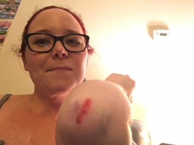
One night last week, I took Astoria out before bed. I had gone out before and checked for skunks before bringing her out. As I was reaching to unlatch the gate, I realized there was a skunk about a foot away from me. I turned to run away from it but Astoria was next to me and I tripped over her. I somehow managed to stay on my feet for about fifteen feet while running at full tilt before finally crashing to the ground on my concrete patio. I hit the ground pretty hard. I managed to open wounds on my left ankle, both knees, both hands, and left elbow. I have a spectacular bruise on my left hip extending down the bruise.

Falling down hurts a lot more than it did ten years ago so I just lay there for a minute. Astoria lay down next to me because she seemed to think this was a bonding exercise. In the split second when I landed on my elbow, my brain decided that I had broken my arm again. I have broken that arm before, twice. Without any evidence at all that anything was seriously wrong with the elbow, my mind had populated a dystopic future for me and my broken arm. I was pretty banged up but there were no serious injuries. I had foreseen the next six weeks of my life dealing with a broken elbow and I basically had a bad scrape that bled. Catastrophizing at its best.

image

In case you are wondering, Astoria did not attack the skunk (a fact that shocks me). When I sat up, the skunk was still standing in the same spot. It had sprayed a tiny bit, so little that it didn’t hit me or Astoria. After a few seconds of staring at me, it turned around and left. I cannot believe we didn’t get sprayed.

People with serious health issues spend a lot of time assuming crash positions and bracing for the pain. We are frequently met with resistance and spend a lot of time having the same conversation with ten people who still don’t rectify the issue.  I am always afraid that today will be the day that something truly awful happens.

But it usually doesn’t. I am learning how to live here, in this space where sometimes things work out.

The price of surviving anaphylaxis

Anaphylaxis is a severe, multisystem allergic event. It is a medical emergency and can be fatal. A 2015 WAO update stated that Americans have a 1.6% risk of anaphylaxis over the course of their lifetime. In the US, anaphylaxis was fatal in 186-225 patients annually, a frequency of 0.63-0.76 per million people. 30-43% of patients with a history of anaphylaxis will have a recurrence.

Epinephrine is the only drug known to decrease the incidence of death from anaphylaxis and should be administered as the first-line agent. Delay in administration of epinephrine has been shown to directly increase the risk of death from anaphylaxis. The use of epinephrine autoinjectors by patients at risk of anaphylaxis is critically important to managing patient safety. Despite this, many patients do not have or do not use their epinephrine autoinjectors.

The use of epinephrine autoinjectors in anaphylaxis has been very well studied. A 2014 paper by Wood and colleagues reported the findings from surveying 35,079 patients. They found that 60% of patients did not have an epinephrine autoinjector on their person when anaphylaxis recurred. Another study by Sanchez found 9-28% of patients carried autoinjectors.

Wood found that 52% of patients with a history of anaphylaxis were never prescribed an autoinjector. In a patient group of 261 with history of proven anaphylaxis, a mere 11% used the autoinjector for their most recent anaphylactic event. Not using epinephrine in the appropriate time frame can have grave consequences. In anaphylaxis patients who progressed to cardiac arrest, 67% did not receive epinephrine within an hour of onset. Among patients who died from anaphylaxis, none of them received epinephrine when the first symptoms presented. Only 14% of fatal anaphylaxis patients were administered epinephrine prior to cardiac arrest.

In many instances, economics is to blame for not carrying an epinephrine autoinjector. A truly stunning statistic is that 50% of anaphylaxis patients do not fill prescriptions for autoinjectors once the cost is over $300. In the previously mentioned 2014 Wood publication, 41% of anaphylaxis patients reported a household income of less than $50,000. Despite being uniformly accepted as a medication used to prevent death and severe complications resulting from anaphylaxis, epinephrine autoinjectors are often not classified by insurance companies as a preventative medication. An analysis of American insurance plans found that the two pack of Epipens was classified as a tier 1 or 2 medication in 67% plans; tier 3 or 4 in 6% plans; “approved” without any contribution to the cost of the drug in 5% of plans; and 22% plans did not cover Epipens at all.

A 2012 paper assessed how patient cost related to adherence to treatment recommended by their provider for a wide array of conditions. Eaddy reported that of the 66 studies evaluated 85% (56 studies) demonstrated that patients were less likely to adhere to the treatment plan when their costs increased.  High out-of-pocket costs were definitively shown to decrease patient use of preventative health care measures, resulting in poorer outcomes.  Increasing copays and deductibles impede access to life saving medication for anaphylaxis patients. If epinephrine autoinjectors were classified universally as preventative medications, insurance companies would be obligated to fully cover the associated costs. They would also be prevented from requiring patients to pay large out of pocket costs for autoinjectors as contribution to deductibles.

Kaplan reported that only 11% of patients with a history of anaphylaxis refill their epinephrine autoinjectors as needed. Instead, many patients rely on expired autoinjectors. Epinephrine is an inherently unstable molecule that degrades quickly when exposed to oxygen or light. A study in 2000 showed that while autoinjectors still functioned as intended up to 90 months after expiration, epinephrine concentration was significantly reduced. Still, expired epinephrine is still better than no epinephrine in the event of anaphylaxis as the benefit would outweigh the risk.

Epinephrine autoinjectors are designed to be stored at 20-25°C but tolerate occasional exposure to higher or lower temperatures in the range of 15-30°C. While heat is known to hasten degradation of epinephrine, freezing apparently is not. The 2015 WAO update mentioned that if autoinjectors are frozen, epinephrine concentration is not affected and that patients can use them as long as they are completely thawed. (I find this really wild, I had never heard of this before.)

Exorbitant costs prevent anaphylaxis patients from having ready access to epinephrine autoinjectors, the only first line medication for anaphylaxis. 50% of patients do not fill prescriptions for epinephrine autoinjectors when their cost is over $300. With the cost of autoinjectors well into the hundreds of dollars for many patients, millions of people in the US may be unable to afford this lifesaving medication for which is there is no alternative.

References:

Simons FER, et al. 2015 update of the evidence base: World Allergy Organization anaphylaxis guidelines. World Allergy Organization Journal 2015: 8(32).

Wood RA, et al. Anaphylaxis in America The prevalence and characteristics of anaphylaxis in the United States. J Allergy Clin Immunol 2014: 133, 461-467.

Noimark L, et al. The use of adrenaline autoinjectors by children and teenagers. Clinical & Experimental Allergy 2012: 42(2), 284-292. Eaddy MT, et al. How patient cost-sharing trends affect adherence and outcomes. Pharmacy & Therapeutics 2012: 37(1), 45-55.

Simons FER, et al. Outdated EpiPen and EpiPen Jr autoinjectors: Past their prime? J Allergy Clin Immunol 2000: 105(5), 1025-1030.

Sanchez J. Anaphylaxis. How often patients carry epinephrine in real life? Rev Alerg Mex 2013: 60, 168-171.

Kaplan MS, et al. Epinephrine autoinjector refill history in an HMO. Curr Allergy Asthma Rep 2011: 11, 65-70.)

Kim JS, et al. Parental use of EpiPen for children with food allergies. J Allergy Clin Immunol 2005: 116(1), 164-168.