Skip to content

November 2014

Food allergy series: Atopy, risk factors and frequency

Food allergy is used widely to describe any adverse reaction to food, but scientifically is considered to include only IgE mediated reactions. For the purposes of this series, I will use this term in the same way. However, there are several other conditions that can cause severe food reactions via different mechanisms and all will be addressed in upcoming posts.

Food allergy causes a reaction to a specific allergen following contact with the skin or mucosa, generally the mouth and GI tract. Colloquially, the terms sensitization and allergy are often used interchangeably, and while they are related, they are not the same. Sensitization is when the body makes allergen specific IgE in the blood or skin. Allergy is sensitization with accompanying symptoms.

Atopy refers to the tendency of a person to develop diseases of allergic nature, like asthma and atopic dermatitis. Having one atopic condition makes a person more likely to develop others. The term “allergic march” has been used to describe the progressive accumulation of atopic conditions from the first year of life through late childhood. Though first described in US patient groups, it has also been observed in several other countries. It typically begins with atopic dermatitis and progresses to include allergic rhinitis, asthma and food allergy. Severe eczema within the first six months of life is associated with increased risk of peanut, milk and egg allergies, for example.

Most food allergies are due to egg, cow’s milk, wheat, soy, shellfish, fish, peanuts and tree nuts. 30.4% of children with food allergy have multiple of them. Kids with multiple food allegies are more likely to have severe reactions, as are adolescents from 14-17 years of age. Peanuts, cashews, walnuts and shellfish allergies are most likely to be severe.

The largest studies on frequency of food allergy use self reported information. These types of surveys are not the gold standard. For example, it has been shown that a large number of people eliminate foods from their diets based on suspicion of allergy. These are often unnecessary. 89% of patients with atopic dermatitis were shown to have no reactions to suspect foods on oral challenge. Still, self reported studies are the largest body of data available.

Out of 38,480 households in the US, 2.4% had children with multiple food allergies and 3% had kids with history of severe allergic reaction. Across 9,667 Canadian households, there was an overall 8% rate of food allergy, in line with findings in the US. Australian studies have focused more specifically on frequency of individual allergens and have used oral food challenges in determining allergy frequency. Out of 2,848 one year old children in Melbourne, 3% were allergic to peanut and 8.9% were allergic to raw egg.

Risk factors for developing food allergies are complicated and will be addressed in greater detail in coming posts. Atopy, low vitamin D, reduced consumption of omega-3 polyunsaturated fatty acids, reduced consumption of antioxidants, increased use of antacids, obesity, increased hygiene, and delaying exposure to common allergens have all been cited as risks. Family history of food allergy as well as specific genetic alleles and HLA profiles have also been tied to food allergies.

In children, males are more likely to develop food allergies, with Black children being the most likely, and Asian children being more likely than White children. Children of immigrants living in the US are at higher risk than children born to American born parents in the US. Food allergy is associated with increased affluence, with people living in households earning more than $50,000/year being more likely to receive a diagnosis. However, it is not clear if this is due to increased access to healthcare.

Childhood food allergies are often thought to be “outgrowable.” Allergies to milk, egg, wheat and soy are more likely to resolve. Peanut, tree nut, fish and shellfish allergies are more likely to persist. Unfortunately, resolution of allergies is on a declining trend.


References:

Sicherer, Scott, Sampson, Hugh. Food allergy: Epidemiology, pathogenesis, diagnosis and treatment. Journal of Allergy and Clinical Immunology. 2014, 133(2): 291-307.

Sicherer, Scott, Leung, Donald. Advances in allergic skin disease, anaphylaxis and hypersensitivity reactions to food, drugs and insects in 2013. Journal of Allergy and Clinical Immunology. 2014, 133(2): 324-334.

Gupta, Ruchi, et al. Childhood food allergies: current diagnosis, treatment and management strategies. Mayo Clinic Proceedings. 2013, 88(5): 512-526.

Inconvenient: The reality of living with food allergies

Earlier this week, a video was circulating on the internet showing a school board meeting in Michigan. In it, school board members were discussing food allergies and the effect they have on foods being allowed in school.

“Well, you should just shoot them,” a school board member said. She threw her arms up and shrugged a little after she said it. She was clearly kidding. It was a joke to her. It is the sort of thing said when forced to discuss inconveniences at length. She said it because food allergies are inconvenient and she was tired of talking about inconvenient things.

Linda Grossman did not want to harm food allergic children. She was just tired of talking about food allergies.

I am also tired. I am tired of people saying things like this that are meant in jest but are insidious and disrespectful and deeply hurtful.

I am tired of people behaving like food allergies are a personality attribute, not a life threatening medical condition.

I am tired of people feeling like they have more of a right to convenience than food allergic people have to be alive.

As of 2010, an estimated 8.96% of adults and 6.53% of children in the US are food allergic. Averaged, this means that about 1/13 people in the US have food allergies. 1/13 means that on average, each classroom in the US can be expected to have 1-2 food allergic students. Among asthmatic children, the frequency of food allergy skyrockets to a whopping 24%. Childhood food allergies cost approximately 25 billion dollars a year in the US.

Frequency of food allergy rose 50% between 1997 and 2011, with peanut allergy alone tripling in that time. As many as 40% of food allergic children have previously had severe anaphylactic reactions that could have resulted in death. Severe reactions requiring hospitalization have increased sevenfold in the last ten years in Europe.

Food allergy is the most common cause of anaphylaxis outside of a hospital setting and results in 200,000 emergency department visits yearly. This equates to one person every three minutes. It is estimated that 100-200 people die from food allergies each year in the US, with numerous conflicting reports.

I am tired of food allergic people and families having to beg for steps to help prevent DEATH.

I am tired of people feeling that making practical changes to prevent people from DYING is inconvenient.

I am tired of people making jokes about a medical issue that KILLS people.

I am tired of food allergic people being excluded from everything because their LIFE THREATENING condition is INCONVENIENT for the other people there.

I am tired of living in a world that prioritizes convenience over the health and welfare of its people.

I am tired of people who should damn well know better spouting dangerously inaccurate facts about food allergy. So I’m putting all my other posts on hold and we’re going to talk about food allergies, FPIES, eosinophilic gastrointestinal disease, celiac disease, and a-gal allergy.

This is not a joke. Anaphylaxis is not a joke. Food allergic people are not jokes. It’s time to stop pretending comments like these aren’t damaging. It’s time for us to stand up and fight.

The other side of the sky

I am the type of person who experiences memory through music. I cannot always remember the vividness of color or feeling when revisiting in my mind, but one verse immerses me in the rich details of sense memories. Three right chords and I am reliving it.

A few days ago, as I was stepping into the shower, Wagon Wheel by Old Crow Medicine Show came on. Immediately, I was in Portland, Oregon last July. I was going for a walk well past midnight, my Epipens in my dress pocket, clanging against my thigh with each step. The air was still warm, humid, but not cloying; just sort of close in the way summer sometimes is. I signed along to the song in ASL as I walked, an old habit from when I was learning.

Driving across the bridge into Portland was the culmination of surviving years of surgeries and shocks and lost pieces of myself. I felt amazing there, filled with this lightness, but also sad around the edges. This disease is so unpredictable. I loved it there so much and I don’t know if I’ll ever get there again.

Last week, I scheduled an appointment with my colorectal surgeon for January to discuss the removal of the end of my GI tract. It no longer has any function and causes me a fair amount of grief. That same day, I booked my flights to Colorado. I was not feeling great emotionally because frankly I’m tired of surgeries and procedures but physically I have been feeling much better. I am sleeping at night and not vomiting every day and don’t feel like a zombie all the time. Anything better than normal feels like such an improvement.

In the next three months, I am planning to visit Florida, Minnesota, California and potentially Hong Kong. It’s a lot of travel for anyone, let alone someone like me. That’s the thing about feeling better – it gives you this artificial bravery to do things you normally wouldn’t. It makes you feel like you can do things you know you can’t. What if I could, though? What if I could do all things I think I can?

I had one of those cries in the shower that night, when Wagon Wheel came on, the kind with wrenching, full body sobs. It had been building all week. Every morning that week I woke up feeling okay and all day I waited for it to get worse. I was afraid of how bad it would feel when this good went away. I was afraid I would remember that I can’t do all these things I want to do, and I was afraid that I would be right.

When I have a really good day, I tell people that I feel like I could fly, like I could touch the sky if I wanted to. Feeling good after not for so long does strange things to your mind. It makes it feel like you can bend the limits of your reality.

Standing in the water, I had an image in my mind of me touching the sky only to have it break apart under my hand. And on the other side of the sky, there was this other place, with no limits, and it terrified me.

When I feel good, part of me is afraid of feeling bad again. But I think there is also a part that is afraid for another reason. I think part of me has no idea how to function outside of these confines my disease has built for me. I think if I was healthy tomorrow, I wouldn’t even remember how to live anymore.


MTHFR, folate metabolism and methylation

MTHFR (methylenetetrahydrofolate reductase) is an enzyme involved in folate metabolism. It is rate limiting, which means that if there is not enough, your body cannot metabolize enough folate; if there is too much, it metabolizes too much. How much folate your body is able to metabolize is directly related to how much MTHFR you have in your body.   Some folate broken down by your body is used to methylate DNA. There has been much conflicting evidence, but it seems that low folate in the body is associated with less DNA methylation overall, which may be associated with cancer (Crider, 2012.)

A single nucleotide polymorphism (SNP) is a change in DNA sequence in which one nucleotide (a DNA building block) is changed. Importantly, SNPs are common. In fact, they are so common that the way your body codes its DNA allows for SNPs to not change gene expression in many cases. This is called wobble. Three DNA building blocks in a row make one amino acid, which is used to build proteins that do things in the body. However, in many cases, the third building block can be any building block and it will still make the same amino acid. Please consider this. SNPs are so common that your cells know that in many cases, 1 in every 3 DNA building blocks can be anything and it won’t change a thing.

SNPs have become the topic of increased interest, both by medical and scientific professions and by lay people. It is certainly true that some SNPs play a role in development and progression of diseases. MTHFR has been found to have up to 24 reported SNPs, with two being of particular interest. These are C677T and A1298C.

The normal (or “wildtype”) form of MTHFR has a cytosine (C) nucleotide where people with the C677T mutation have a thymine nucleotide. If you have two copies of the regular gene, you are 677CC and homozygous for the regular form of the gene (the allele.) If you are 677CT, you have one copy of the allele with the SNP. If you are 677TT, you have two copies of the allele with the SNP. About 10% of North Americans are 677TT, meaning they have two copies of the mutated allele. It is most common in Hispanics and those of Mediterranean descent, next most common in Caucasians and least common in African Americans (Schneider, 1998.)

Being homozygous for 677TT can cause a mild MTHFR deficiency because that this form of the enzyme is generally less stable, which means it breaks down faster than usual. People with this profile are also more likely to develop mild hyperhomocysteinemia, an elevation of homocysteine in the blood. Homocysteine is consumed in the metabolism of folate, and because there is less MTHFR with the 677TT mutation, the homocysteine is not all getting used. It is also elevated in cases of B6, B12 and folic acid deficiency.

Increased homocysteine has been studied for its possible relationship to health issues, including increased clotting, strokes, schizophrenia and osteoporosis. Despite multiple studies (to be honest, quite a lot of studies), the results are really non-uniform. Because it was linked earlier to cardiovascular disease, multiple studies investigated the benefits of lowering homocysteine. In diabetic nephropathy patients, treating to lower homocysteine actually doubled cardiovascular events including heart attack and stroke, some leading to death, as well as decreased renal function (House, 2010.) But another study found lowering homocysteine decreased the risk of stroke by as much as 25% (Lonn, 2006.)

The effects of the C677T SNP have been most well studied in regards to development of cancers. Folic acid is a key metabolite in the development and proliferation of cells, so deficiency can be limiting to cell division. 677TT patients who are not folate deficient are actually 50% less likely to develop colorectal cancer or colorectal adenoma (Chen, 1999.) 677TT patients who are folate deficient have at least the same risk as 677CC patients and potentially more risk (Slattery, 1999.) 677TT patients are over four times less likely to develop acute lymphocytic leukemia. They have the same risk of developing acute myeloid leukemia (Skibola, 1999.) Some studies found them to have increased risk of cervical neoplasia, breast cancer, endometrial cancer and gastric cancer, but all of these studies were done on small populations (Xia, 2014.)

The other MTHFR SNP that gets a lot of attention is A1298C. At position 1298 of the MTHFR gene, the wildtype allele has adenine. In some people, it is substituted for cytosine. Wildtype is homozygous for adenine there and they are called 1298AA. If you have one copy of the SNP, you are 1298AC. If you have two copies, you are 1298CC. The A1298C has much less effect on the stability of the MTHFR protein than the C677T mutation. It is not known to cause elevation of homocysteine levels. There has been a lot of controversy over whether this mutation can cause a deficiency of BH4, tetrahydrobiopterin. BH4 is important in formation of neurotransmitters and nitric oxide, as well as consuming ammonia. Low levels of BH4 have been tied to phenylketonuria and trials with BH4 supplementation have seen encouraging results (Michals-Matalon, 2007.)

In the last six months or so, I have read about fifty scientific papers on MTHFR or related topics. I did this because I originally planned an MTHFR post for last summer. I didn’t do a post because the data is a mess. You cannot ascertain much of use from the peer reviewed literature on the C677T and A1298C mutations – and not for lack of effort. These mutations have been very well studied.

MTHFR mutations and methylation are talked about a lot in the mast cell community. Many people believe that having an MTHFR mutation severely impacts folate metabolism, which in turn means there is not enough methylation, and this dysregulation causes overexpression of genes causing disease. I have searched thoroughly for a link. Really thoroughly. I cannot find any link that is not the idea of one person and researched by that one person, usually outside of peer reviewed settings. I cannot find any link that is not described in detail by a person who does not stand to gain financially from patients who share their beliefs. I am not saying that MTHFR is definitely not linked to mast cell disease. I’m saying I can’t find any proof that it is. I can’t even find anything that SUGGESTS that it is. Might people with these mutations feel better with appropriate folic acid supplementation? Probably. Is that the same thing as causing mast cell disease? Certainly not. It is certainly not the same thing.

I think personal stories hold a lot of power. If your personal story is that your mast cell disease is significantly better controlled by addressing your MTHFR mutation, then I think that is fantastic. I think it is entirely possible that this is the case for many. But I do not believe it causes mast cell disease. And I think everyone would feel better with appropriate levels of folate, as it is an important player in many vital reactions.


References:

Schneider JA, Rees DC, Liu YT, Clegg JB (May 1998). “Worldwide distribution of a common methylenetetrahydrofolate reductase mutation”. Am. J. Hum. Genet. 62 (5): 1258–60

M L Slattery, et al. Methylenetetrahydrofolate reductase, diet and risk of colon cancer. Cancer Epidemiol Biomarkers Prev., 8 (1999), PP 560S-564S.

House, AA; Eliasziw, M; Cattran, DC; Churchill, DN; Oliver, MJ; Fine, A; Dresser, GK; Spence, JD (Apr 28, 2010). “Effect of B-vitamin therapy on progression of diabetic nephropathy: a randomized controlled trial.”. JAMA: the Journal of the American Medical Association 303 (16): 1603–9.

Lonn, E; Yusuf, S; Arnold, MJ; Sheridan, P; Pogue, J; Micks, M; McQueen, MJ; Probstfield, J; Fodor, G; Held, C; Genest J, Jr; Heart Outcomes Prevention Evaluation (HOPE) 2, Investigators (Apr 13, 2006). “Homocysteine lowering with folic acid and B vitamins in vascular disease.”. The New England Journal of Medicine 354 (15): 1567–77

Skibola CF, Smith MT, Kane E, Roman E, Rollinson S, Cartwright RA, Morgan G’ (October 1999). “Polymorphisms in the methylenetetrahydrofolate reductase gene are associated with susceptibility to acute leukemia in adults”. Proc. Natl. Acad. Sci. U.S.A. 96 (22): 12810–5.

Kim, Y I, et al. Methylenetetrahydrofolate reductase polymorphisms, folate and cancer risk: a paradigm of gene-nutrient interactions in carcinogenesis. Nutr Rev 58 (2000), pp 205-209.

Crider, Krista, et al. Folate and DNA Methylation: A Review of Molecular Mechanisms and the Evidence for Folate’s Role. Adv Nutr January 2012 Adv Nutr vol. 3: 21-38, 2012

Xia, Lei-Zhou, et al. Methylenetetrahydrofolate reductase C677T and A1298C polymorphisms and gastric cancer susceptibility. World J Gastroenterol. Aug 28, 2014; 20(32): 11429–11438.

J Chen, et al. MTHFR polymorphism, methyl-replete diets and the risk of colorectal carcinoma and adenoma among US men and women: an example of gene–environment interactions in colorectal tumorigenesis. J. Nutr., 129 (1999), pp. 560S–564S

DNA Methylation: How it works

DNA methylation is one of the ways your cells control which genes to express. It is an example of epigenetic modification. Epigenetics mechanisms like this do not change the DNA sequence, only the way the genes are expressed. Whether or not DNA methylation is heritable is not clear.

This is how DNA methylation works:

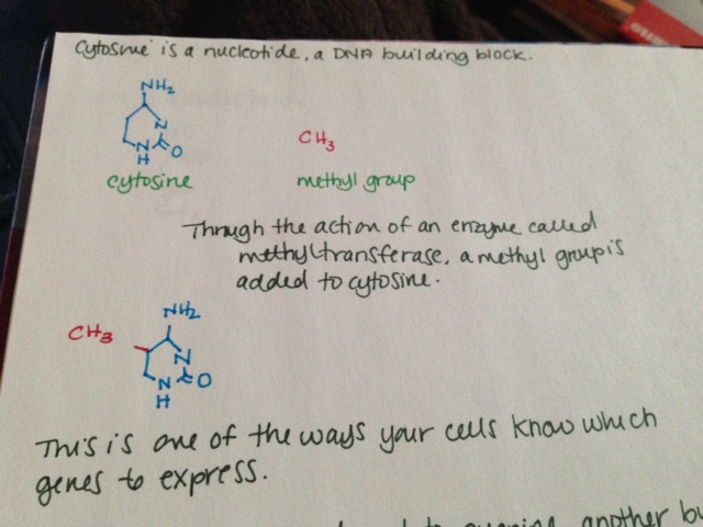
  • Cytosine is a nucleotide, a DNA building block.
  • Through the action of an enzyme called methyltransferase, a methyl group is added to cytosine.
  • This is one of the ways your cells know which genes to express.
  • Cytosine is often found next to guanine, another building block. This is sometimes shown in literature as “CG.”
  • Cytosine and guanine are connected by a phosphate group. This is sometimes shown in literature as “CpG.”
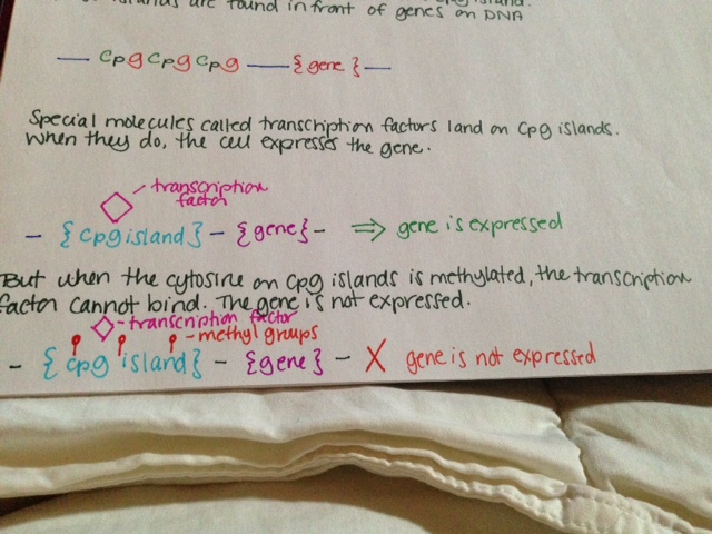
  • A bunch of CpG sites together is called a CpG island. These islands are found in front of genes on DNA.
  • Special molecules called transcription factors land on CpG islands. When they do, the cell expresses the gene.
  • But when the cytosine on CpG islands is methylated, the transcription factor cannot bind. The gene is not expressed.

See pictures below.

Methylation is known to have an important role in cancer biology. Methylation of tumor suppressor genes causes the tumor suppressors not to be expressed, resulting in cancer.

Methylation 1Methylation 2Methylation 3

Lesser known mast cell mediators (Part 4)

Interleukin-1a (IL-1a) is largely responsible for inflammation, fever and sepsis. It activates TNF-a and the work very closely together. Their cofunctions include PGE2 synthesis, nitric oxide production, insulin resistance and IL-8 and chemokine production.

Interleukin-1b (IL-1b) has been implicated in several autoinflammatory syndromes. It is also important in cell proliferation, differentiation and apoptosis. Its induction of COX2 cytokine in the nervous system contributes to inflammatory pain hypersensitivity.

Interleukin-2 (IL-2) is crucial in prevention of autoimmune disease by regulating T cell differentiation. It is also thought to be involved in itchiness and psoriasis. IL-2 is used in the treatment of cancers.

Interleukin 3 (IL-3) drives the differentiation of multipotent hematopoietic stem cells into myeloid progenitor cells. If IL-7 is also present, they can work synergistically to trigger differentiation into lymphoid progenitor cells. IL-3 induces proliferation of all myeloid cells (including mast cells) along with other cytokines like IL-6. It supports growth and differentiation of T cells from bone marrow when an immune response is triggered.

Interleukin 4 (IL-4) changes naïve T cells to T helper cells, which secrete chemicals to drive actions of other immune cells. T helper cells then secrete additional IL-4 to perpetuate the cycle. IL-4 participates in the airway inflammation seen in allergic asthma.

Interleukin 5 (IL-5) encourages growth of B cells and antibody secretion as well as eosinophil activation. It is heavily involved in allergic diseases, particularly those in which eosinophils are notably increased. Mepolizumab is a monoclonal antibody against IL-5 that can reduce excessive eosinophils.

Interleukin 6 (IL-6) mediates fever and the acute phase inflammatory response. It is secreted to stimulate bone resorption and inhibitors of IL-6 are used to treat osteoporosis (including estrogen.) It inhibits TNF-a and IL-1. Unusually, it also has anti-inflammatory behaviors, particularly during exercise in the muscle.

Interleukin 9 (IL-9) increases cell proliferation and impedes apoptosis, cell death, of hematopoietic cells. It is particularly important in asthma and bronchial hyperresponsiveness.

Interleukin 10 (IL-10) is an anti-inflammatory molecule involved in regulating the JAK-STAT pathway. It counteracts many of the inflammatory effects of mast cells, often by interfering with production of substances like interferons and TNF-a.   Exercise increases levels of this molecule.

Interleukin 13 (IL-13) is critical in initiation of airway disease. It induces matrix metalloproteinases to act. IL-13 can also induce IgE release from B cells. It is effectively a link between allergic inflammatory cells and the non-immune cells they interact with. Excessive , IL-13 causes airway hyperresponsiveness, goblet cell metaplasia and oversecretion of mucus.


I think I might have mast cell disease: FAQ

What kinds of symptoms do mast cell patients have?

Mast cell disease can cause a variety of symptoms. Each person has their own unique constellation of complaints, and they can vary from day to day. Mast cell patients often have allergic type reactions to many things. They may have had anaphylaxis in the past, but that is not always the case.

What kind of doctor should I see if I think I have mast cell disease?

Due to the fact that mast cell disease can affect multiple body systems, it is managed by doctors of multiple disciplines. Immunologists, dermatologists, gastroenterologists and hematologists/ oncologists all may treat mast cell disease. It really depends who is familiar with mast cell disease in your areas. Immunologists are often the first stop for patients investigating mast cell disease.

Will any doctor know about mast cell disease?

No. Mast cell disease is uncommon. Many doctors are only aware of the types associated with pathologic rashes (cutaneous mastocytosis) or proliferation of mast cells in the bone marrow (systemic mastocytosis.)

How do I get determine if I have mast cell disease?

Labs for diagnosing mast cell disease include serum tryptase (a blood test), n-methylhistamine (24 hour urine test) and D2/F2a prostaglandin (24 hour urine test.) These tests are time sensitive for many patients and have special handling in most labs. Depending on these results, a bone marrow biopsy may be needed.

Can I have mast cell disease if my tryptase is normal?

Yes. 15% of patients with systemic mastocytosis have normal tryptase levels, and the majority of MCAS patients have normal tryptase levels.

How is mast cell disease treated?

Treatment generally focuses on the symptoms. The most common treatments include antihistamines, leukotriene inhibitors and mast cell stabilizers.

Will I feel better with treatment?

Most people feel better with treatment than without, but how much each person recovers is individual. Lifestyle modifications and medications can help many people live a full life.

Is mast cell disease curable?

No. Patients may have a remission from symptoms, but they will always have mast cell disease.

Symptoms of mast cell disease

The following is a generalized list of common symptoms associated with mast cell disease. It is not comprehensive and does not include laboratory or associated diagnoses.

General: fatigue, malaise (“being out of it”), weakness, severe unprovoked sweating, weight gain or loss

Skin: Rashes and lesions of any kind, itching, flushing, angioedema, stretch marks, dermatographism, poor wound healing, alopecia, abnormalities of finger or toenails

Eyes: Irritated eyes, red eyes, excessive tearing, dry eyes, difficulty focusing vision, lid tremor, sensitivity to lightness, including sunlight, general inflammation

Ears: Inflammation of the ear, “ear infections,” hearing loss, sensitivity to sound, ringing in the ears

Mouth/throat/sinuses: Generalized pain of several qualities, but often burning, ulceration, “canker” sores, swelling (angioedema), dental decay, abnormalities in taste, taste of metal, throat discomfort or irritation, need to clear throat frequently, post nasal drip, nose bleeds, irritation of sinuses, sinus congestion

Respiratory: laryngitis, bronchitis, pneumonitis (frequently confused with pneumonia), recurrent cough (usually dry), shortness of breath, wheezing

Cardiovascular: lightheadedness, weakness, dizziness, vertigo, fainting, high or low blood pressure, palpitations, rapid heartbeat, abnormalities in heart rhythm, chest pain, hemorrhoids, edema

Gastrointestinal: abdominal pain, diarrhea, constipation, difficulty swallowing, swelling of any part of the GI tract, nausea

Neurologic: Headache, migraine, “about to faint,” fainting, numbness, pins and needles, neuropathy, tics, tremors, seizures

Psychiatric: Anger, depression, PTSD, anxiety, memory difficulties, anxiety, panic disorders, insomnia, sleep disorders

The hallmark of mast cell disease is allergic-type reaction to a variety of stimuli. These can also occur to substances benign to most people, including scents, “hypoallergenic” materials, heat, sunlight and water. Some people experience anaphylactic reactions that require epinephrine.

References:

Afrin, Lawrence B. Presentation, diagnosis and management of mast cell activation syndrome. 2013. Mast cells.

The other kind of hope

I am an optimistic person. My optimism borders on religious; when I despair, it is all that I have. I am good at finding silver linings, at genuinely feeling fortunate or lucky or grateful for the little upturns of bad situations. I enjoy talking to my father when he has to drive me to work. I like snuggling with Astoria when I have to spend the day in bed. I am grateful for my awesome friends, family and coworkers who help me out. All of these things happen because I am sick, manifestations of the impact my illness has on my life. They give me hope that I can keep doing this.

But there is this other kind of hope, more insidious and malignant. I woke up this morning on my own after sleeping for nine hours. This is the second night in a row I have done this. I felt okay when I woke up. Some bone pain, but overall, better than normal. And then it happened, that dangerous optimism – maybe I’m getting better. Maybe this is when I start to get better.

It never is. I know logically that two days of good sleep doesn’t mean I’m headed for a remission. I wish I didn’t feel these things so intensely. But I do.

Even after all this time, I still can’t believe that I will never get better. I can know it in my mind, but my heart just won’t accept that this is anything but temporary. This hope for impermanence can be so painful.

Sometimes I wish I weren’t so hopeful. It is just so hard to live with the perpetual disappointment.

Lesser known mast cell mediators (Part 3)

Substance P is a neurotransmitter and modulates neurologic responses. It is found in many sensory nerves as well as the brain and spinal cord. It participates in inflammatory responses and is important in pain perception. It is involved in mood disorders, anxiety, stress, nerve growth, respiration, neurotoxicity, nausea, vomiting and pain perception. Its release from nerve fibers into the skin, muscle and joints is thought to cause neurogenic inflammation.

Urocortin is related to corticotropin releasing factor (CRF.) It strongly suppresses blood pressure and increases coronary blood flow. It is thought to have a role in increasing appetite during times of stress.

VEGF-A (vascular endothelial growth factor A) is a member of the platelet derived growth factor (PDGF)/VEGF family. It is important in nerve biology and is the substance mainly involved in inducing growth of blood vessels. It is heavily involved in diseases that involve blood vessels, like diabetic retinopathy and macular degeneration. It is a vasodilator and increases permeability of the smaller vessels.

VIP (vasoactive intestinal peptide) is a small protein like molecule used by nerve cells for communication. It stimulates heart contraction, vasodilation, lowers blood pressure, and relaxes the smooth muscles of the trachea, stomach and gall bladder. It also inhibits gastric acid secretion and absorption in the intestine.

Mast cell kininogenase removes a portion of a compound to release active bradykinin. This is important in the kinin system.

Phospholipase A2 promotes inflammation by initiating formation of arachidonic acid, the precursor needed to form many inflammatory molecules, including prostaglandins. Excessive levels of phospholipase A2 can lead to increased vascular inflammation, such as a seen in coronary artery disease and acute coronary syndrome. Elevated PLA2 is found in the cerebrospinal fluid of people with Alzheimer’s disease and multiple sclerosis.

Corticotropin releasing hormone (CRH) is a hormone and neurotransmitter. High CRH levels have been associated with Alzheimer’s disease and severe depression. CRH is produced in the hypothalamus and is carried to the pituitary gland, where it stimulates secretion of adrenocorticotropic hormone (ACTH.) ACTH drives synthesis of cortisol and other steroids. Imbalance of these hormones can have dire consequences.

Endothelin is the most potent vasoconstrictor currently described. It raises blood pressure and if uncontrolled, hypertension may result. It is involved in many disease processes, including cardiac hypertrophy, type II diabetes and Hirschsprung disease.

Chondroitin is found largely in connective tissues and is a principal component of cartilage. It is typically bound to other components when released from mast cells and interacts with a variety of molecules.

Hyaluronic acid is widely found in epithelial, neural and connective tissues. It participates in a variety of reactions and sees significant turnover daily. When hyaluronic acid is degraded as part of the turnover, its degradation products can cause inflammatory responses.