Skip to content

Lisa Klimas

I'm a 35 year old microbiologist and molecular biologist with systemic mastocytosis, Ehlers Danlos Syndrome, Postural Orthostatic Tachycardia Syndrome, Adrenal Insufficiency, and an assortment of other chronic health issues. My life is pretty much a blast.

Supporting materials for explaining mast cell disease to non-health care professionals

A patient asked how to explain mast cell disease at a high level in a short period of time to legal professionals outside of medical field. The information presented here is largely taken from several sources, materials I had previously prepared for several cases to explain mast cell disease to non-health care professionals. It was difficult to organize all the concepts in the manner I prefer without rewriting everything. For this reason, the style of this article is more “stream of consciousness” than my typical writing.

The rest of the information discussed here is taken directly from work presented on MastAttack.

I also included technical details in appendices for reference. This information is also derived from articles published on MastAttack.

****

Mast cell activation disorders (MCAD) are a group of conditions in which the mast cells in the body do not function correctly. Mast cells are responsible for allergic responses. They are the allergy cells in tissues.

In MCAD, patients can have allergic type reactions to things they are not allergic to, called mast cell reactions or degranulation reactions. These reactions can be very severe and even life threatening.

Mast cell reactions are caused by mast cells being improperly activated. Triggers vary from person to person. More common triggers include heat, cold, friction (especially on the skin), sunlight, foodstuffs, physical exertion, stress, dyes and fragrances. Because mast cell reactions can be dangerous, trigger avoidance is a crucial component of managing mast cell disease.

These reactions vary from person to person. Symptoms can include, but are not limited to, nausea, vomiting, hives, rashes, itching, flushing (turning red), dizziness, confusion and irritability. Symptoms are caused by the chemicals released by the mast cells. In severe cases, mast cell reactions can culminate in anaphylaxis, a severe, life threatening allergic reaction.

Due to the ubiquity of mast cells in bodily tissues, mast cell disease has the potential to cause an array of symptoms across multiple systems. Examples include flushing; acute or chronic urticaria; brain fog; anxiety; angioedema; tachycardia; hypotension;  diarrhea; and chronic GI dysmotility, among others.

While many providers believe that hypertension excludes both mast cell disease and anaphylaxis, hypertension is common in mast cell patients. Data described and published in 2016 demonstrate that hypertension could affect as many as 31% of mast cell patients, including patients with systemic mastocytosis and mast cell activation syndrome.

In my experience, gastrointestinal symptoms are frequently the most debilitating and, in some instances, the most dangerous.

Mast cell disease greatly increases the risk of anaphylaxis. Patients are advised to carry two epinephrine autoinjectors at all times.

Allergic reactions to food are overwhelmingly common in this population. Furthermore, food reactions due to mast cell activation exhibit atypical features that complicate diagnosis and management. It is not unusual for mast cell patients to react to foods or medications they typically tolerate when mast cell activation is increased. Regaining tolerance for these substances can be complicated and time consuming. Regaining tolerance is not always possible.

Mast cell food reactions are influenced by the level of histamine circulating in the body at the time of consumption. Histamine levels fluctuate throughout the day in everyone as mast cell activation is necessary to perform many appropriate actions such as digestion and regulation of sleep. Other healthy activities, such as exercise or use of certain medications, also raise the level of circulating histamine. For these reasons, the severity of mast cell food reactions can fluctuate throughout the day.

Histamine level is also impacted by acute health state. For example, due to the importance of mast cell activation in immune defense, patients may find that their mast cell food reactions are more severe while fighting an infection, recovering from surgery, or healing a wound.

Given the nutritional constraints of limited diets, many patients eventually require use of elemental formulas, or partial or total parenteral nutrition (TPN) given intravenously.

Diagnosis

Mast cell activation syndrome is recently described phenomenon. As such, there are multiple sets of diagnostic criteria that reflect the personal experience of providers with mast cell patients. All sets of criteria include the following three items: recurrent or chronic symptoms of mast cell activation; objective evidence of excessive mast cell mediator release; and positive response to medications that inhibit action of mast cell mediators.

Mast cells produce and release a number of sensitive mediators that can be quantified and may be elevated in mast cell patients. Please note that testing is complicated as mediator levels can fluctuate wildly for a number of reasons. Elevation of serum tryptase; histamine or its metabolite, n-methylhistamine; prostaglandin D2 or its metabolite 9a,11b prostaglandin F2a; and leukotriene E4 is a positive marker for mast cell activation syndrome.

For further details, please refer to Appendix A: Diagnosis of mast cell activation syndrome and Appendix B: Mediator testing.

Management of mast cell disease

Management of symptoms of mast cell activation is complex and individualized. Typically, mast cell patients take baseline medications, including a second generation H1 antihistamine, like cetirizine; an H2 antihistamine, like famotidine; a mast cell stabilizer, like cromolyn; and a leukotriene inhibitor, like montelukast.

For further details, please refer to Appendix C: Management of mast cell activation syndrome.

 Identification of triggers and causes

Mast cells are most well known for their importance in allergies and anaphylaxis. There are multiple mechanisms through which an allergy develops and affects the body. The most common pathway is via allergen specific IgE. IgE overwhelmingly drives the reaction to common food and environmental allergies as seen in the general population. IgE is also responsible for unusual presentations such as alpha-gal allergy, in which a patient develops an allergy to meat following a tick bite.

While IgE allergies are the most common presentation, there are a number of allergic conditions caused by alternative mechanisms. Examples of allergic GI conditions effected by alternative means include food protein induced enterocolitis syndrome (FPIES); food protein induced allergic proctocolitis; eosinophilic gastrointestinal disease (EGID); oral allergy syndrome; celiac disease; Heiner syndrome; and mast cell diseases, including systemic mastocytosis, mast cell activation syndrome, and monoclonal mast cell activation syndrome.

The current gold standard for identifying IgE mediated allergy triggers in the general population is scratch testing and RAST testing. These tests will not reveal mast cell triggers.

Scratch testing is unreproducible due increased activation of mast cells in the skin.

Scratch testing requires cessation of antihistamines in the days leading up to the test. Mast cell patients are advised not to stop any baseline meds without buy-in from their mast cell specialist for safety reasons.

RAST testing assesses the presence of allergen specific IgE. As most mast cell reactions are not IgE mediated, RAST testing will not identify most mast cell triggers.

If a patient has IgE allergies and mast cell disease concurrently, RAST testing will only identify IgE allergies and will not identify mast cell triggers. The only reliable method for identifying mast cell triggers is food trials. As mast cell reactions can culminate in anaphylaxis, food trials can pose significant risk to the patient.

Due to the fact that mast cell reactions are not mediated by IgE, it is impossible to infer other triggers based upon the structure of the allergen. For example, in the general population, allergy to one opiate increases the risk of allergy to another due to structural similarities. However, mast cell reaction to one opiate does not imply that other opiates would trigger a reaction in the same patient.

While in some instances the ensuing mast cell reaction from a trigger is not life threatening, there is always a possibility that this reaction can culminate in anaphylaxis. Anaphylaxis is a severe, life threatening, multisystem allergic event capable of causing shock, respiratory and cardiac arrest, and death.

****

For additional reading in lay terms, The Mast Cell Disease Fact Sheet is a great resource. The 107 series presents detailed information on a variety of topics in lay terms as well.

For detailed technical information in medical jargon, The Provider Primer Series can be helpful.

****

Appendix A: Diagnosis of mast cell activation syndrome

Diagnostic criteria

  • MCAS is a recently described diagnosis. In the absence of large studies, several groups have developed their own, sometimes conflicting, diagnostic criteria.
  • Differential diagnoses with potential to cause similar symptoms should be considered and excluded[iii].
  • The criteria most frequently used include those by a 2010 paper by Akin, Valent and Metcalfe[iii]; a 2011 paper by Molderings, Afrin and colleagues[iv]; and a 2013 paper by Castells and colleagues[v].
  • The criteria described in the 2011 paper by Molderings, Afrin and colleagues have been updated to include response to medication[vi].
  • Of note, a 2012 consensus proposal[x] was authored by a number of mast cell experts including Valent, Escribano, Castells, Akin and Metcalfe. It sees little practical use and is not generally accepted in the community.
  • The major sets of criteria listed above all include the following features:
  • Recurrent or chronic symptoms of mast cell activation
  • Objective evidence of excessive mast cell mediator release
  • Positive response to medications that inhibit action of mast cell mediators
  • Valent warns that in some cases, patients may not fulfill all criteria but still warrant treatment: “In many cases, only two or even one of these three criteria can be documented. In the case of typical symptoms, the provisional diagnosis of ‘possibly MCA/MCAS can be established, and in acute cases, immediate treatment should be introduced.”[vii]

Evidence of mediator release

  • Mast cells produce a multitude of mediators including tryptase, histamine, prostaglandin D2, and leukotrienes C4, D4 and E4[viii].
  • Serum tryptase and 24 hour urine testing for n-methylhistamine, prostaglandin D2, prostaglandin 9a,11b-prostaglandin F2a; are frequently included in testing guidelines in literature (Castells 2013)[ix], (Akin 2010)[x], (Valent 2012)[xi].
  • It can be helpful to test for other mast cell mediators including 24 hour urine testing for leukotriene E4[xii].

Symptoms associated with mast cell activation

  • Mediator release causes a wide array of symptoms, including hypertension[xv], hypotension, hypertension, wheezing, itching, flushing, tachycardia, nausea, vomiting, diarrhea, constipation, headache, angioedema, fatigue, and neurologic symptoms[iv].
  • In a small MCAS cohort (18 patients), 17% had a history of anaphylaxis[xvii] . A larger data set is desirable.
  • Patients with history of anaphylaxis should be prescribed epinephrine autoinjectors[v]. If patient must be on a beta blocker, they should be prescribed a glucagon injector for use in the event of anaphylaxis[v].

Response to medications that inhibit action of mast cell mediators

  • Treatment of MCAS is complex and may require a number of medications. Second generation H1 antihistamines; H2 antihistamines; and mast cell stabilizers are mainstays of treatment[xvi].
  • Additional options include aspirin; anti-IgE; leukotriene blocker; and corticosteroids[xiii] .
  • First generation H1 antihistamines may be used for breakthrough symptoms[xiii] .
  • “An important point is that many different mediators may be involved in MCA-related symptoms so that the final conclusion the patient is not responding to antimediator therapy should only be drawn after having applied several different antimediator-type drugs[xiii] .
  • Inactive ingredients are often to blame for reaction to mast cell mediator focused medications. Many mast cell patients see benefit from having medications compounded[xvii].

References:

[i] Frieri M, et al. (2013). Mast cell activation syndrome: a review. Current Allergy and Asthma Reports, 13(1), 27-32.

[ii] Molderings GJ, et al. (2013). Familial occurrence of systemic mast cell activation disease. PLoS One, 8, e76241-24098785

[iii] Akin C, et al. (2010). Mast cell activation syndrome: proposed diagnostic criteria. J Allergy Clin Immunol, 126(6), 1099-1104.e4

[iv] Molderings GJ, et al. (2011). Mast cell activation disease: a concise practical guide for diagnostic workup and therapeutic options. Journal of Hematology & Oncology, 4(10), 10.1186/1756-8722-4-10

[v] Castells M, et al. (2013). Expanding spectrum of mast cell activation disorders: monoclonal and idiopathic mast cell activation syndromes. Clin Ther, 35(5), 548-562.

[vi] Afrin LB, et al. (2016). Often seen, rarely recognized: mast cell activation disease – a guide to diagnosis and therapeutic options. Annals of Medicine, 48(3).

[vii] Valent P. (2013). Mast cell activation syndromes: definition and classification. European Journal of Allergy and Clinical Immunology, 68(4), 417-424.

[viii] Theoharides TC, et al. (2012). Mast cells and inflammation. Biochimica et Biophysica Acta (BBA) – Molecular Basis of Disease, 1822(1), 21-33.

[ix] Picard M, et al. (2013). Expanding spectrum of mast cell activation disorders: monoclonal and idiopathic mast cell activation syndromes. Clinical Therapeutics, 35(5), 548-562.

[x] Akin C, et al. (2010). Mast cell activation syndrome: proposed diagnostic criteria. J Allergy Clin Immunol, 126(6), 1099-1104.e4

[xi] Valent P, et al. (2012). Definitions, criteria and global classification of mast cell disorders with special reference to mast cell activation syndromes: a consensus proposal. Int Arch Allergy Immunol, 157(3), 215-225.

[xii] Lueke AJ, et al. (2016). Analytical and clinical validation of an LC-MS/MS method for urine leukotriene E4: a marker of systemic mastocytosis. Clin Biochem, 49(13-14), 979-982.

[xiii] Vysniauskaite M, et al. (2015). Determination of plasma heparin level improves identification of systemic mast cell activation disease. PLoS One, 10(4), e0124912

[xiv] Zenker N, Afrin LB. (2015). Utilities of various mast cell mediators in diagnosing mast cell activation syndrome. Blood, 126(5174).

[xv] Shibao C, et al. (2005). Hyperadrenergic postural tachycardia syndrome in mast cell activation disorders. Hypertension, 45(3), 385-390.

[xvi] Cardet JC, et al. (2013). Immunology and clinical manifestations of non-clonal mast cell activation syndrome. Curr Allergy Asthma Rep, 13(1), 10-18.

[xvii] Afrin LB. “Presentation, diagnosis and management of mast cell activation syndrome.” In: Mast Cells. Edited by David B. Murray, Nova cience Publishers, Inc., 2013, 155-232.

[xviii] Hamilton MJ, et al. (2011). Mast cell activation syndrome: a newly recognized disorder with systemic clinical manifestations. Journal of Allergy and Clinical Immunology, 128(1), 147-152.e2

Appendix B: Mediator testing

Tryptase

  • Tryptase is extremely specific for mast cell activation in the absence of hematologic malignancy or advanced kidney disease. Of note, rheumatoid factor can cause false elevation of tryptase[ix].
  • Serum tryptase levels peak 15-120 minutes after release with an estimated half-life of two hours[vi].
  • Per key opinion leaders, tryptase levels should be drawn 15 minutes to 4 hours after onset of anaphylaxis or activation event (Castells 2013[ii]), (Akin 2010[iii]), (Valent 2012)[iv]). Phadia, the manufacturer of the ImmunoCap® test to quantify tryptase, recommends that blood be drawn 15 minutes to 3 hours after event onset[vii].
  • Serum tryptase >11.4 ng/mL is elevated[i].
  • An increase in serum tryptase level during an event by 20% + 2 ng/mL above patient baseline is often accepted as evidence of mast cell activation[v],[i].
  • Absent elevation of tryptase level from baseline during an event does not exclude mast cell activation[viii].
  • Sensitivity for serum tryptase assay in MCAS patients was assessed as 10% in a 2014 paper[ix].
  • A recent retrospective study of almost 200 patients found serum tryptase was elevated in 8.8% of MCAS patients[x].

Histamine and degradation product n-methylhistamine

  • N-methylhistamine is the breakdown product of histamine.
  • Histamine is degraded quickly. Samples should be drawn within 15 minutes of episode onset[vii].
  • Serum histamine levels peak 5 minutes after release and return to baseline in 15-30 minutes[vii].
  • Sample (urine or serum) must be kept chilled[xi].
  • A recent retrospective study of almost 200 patients found that n-methylhistamine was elevated in 7.4% of MCAS patients in random spot urine and 5.4% in 24-hour urine[xi].
  • Sensitivity of 24-hour n-methylhistamine for MCAS was assessed as 22% in 24-hour urine[ix].
  • Plasma histamine was elevated in 29.3% of MCAS patients[xi].

Prostaglandin D2 and degradation product 9a,11b-prostaglandin F2a

  • 9a,11b-prostaglandin F2a is the breakdown product of prostaglandin D2.
  • Prostaglandin D2 is only produced in large quantities by mast cells. Basophils, eosinophils and other cells produce minute amounts[ix].
  • A recent retrospective study of almost 200 patients found that PGD2 was elevated in 9.8% of MCAS patients in random spot urines and 38.3% in 24-hour urine[xi].
  • PGD2 was elevated in 13.2% of MCAS patients in plasma[xi].
  • 9a,11b-prostaglandin F2a was elevated in 36.8% in 24-hour urine[xi].
  • Prostaglandins are thermolabile and begin to break down in a minutes. This can contribute to false negative results[xi].

Leukotriene E4

  • Leukotriene E4 is produced by mast cells and several other cell types[ix] including eosinophils, basophils and macrophages.
  • A recent retrospective study of almost 200 patients found that LTE4 was elevated in 4.4 % of MCAS patients in random spot urines and 8.3% in 24-hour urine[xi].

References:

[i] Theoharides TC, et al. (2012). Mast cells and inflammation. Biochimica et Biophysica Acta (BBA) – Molecular Basis of Disease, 1822(1), 21-33.

[ii] Picard M, et al. (2013). Expanding spectrum of mast cell activation disorders: monoclonal and idiopathic mast cell activation syndromes. Clinical Therapeutics, 35(5), 548-562.

[iii] Akin C, et al. (2010). Mast cell activation syndrome: proposed diagnostic criteria. J Allergy Clin Immunol, 126(6), 1099-1104.e4

[iv] Valent P, et al. (2012). Definitions, criteria and global classification of mast cell disorders with special reference to mast cell activation syndromes: a consensus proposal. Int Arch Allergy Immunol, 157(3), 215-225.

[v] Lueke AJ, et al. (2016). Analytical and clinical validation of an LC-MS/MS method for urine leukotriene E4: a marker of systemic mastocytosis. Clin Biochem, 49(13-14), 979-982.

[vi] Payne V, Kam PCA. (2004). Mast cell tryptase: a review of its physiology and clinical significance. Anaesthesia, 59(7), 695-703.

[vii] Phadia AB. ImmunoCAP® Tryptase in anaphylaxis. Retrieved from: http://www.phadia.com/Global/Market%20Companies/Sweden/Best%C3%A4ll%20information/Filer%20(pdf)/ImmunoCAP_Tryptase_anafylaxi.pdf

[viii] Sprung J, et al. (2015). Presence or absence of elevated acute total serum tryptase by itself is not a definitive marker for an allergic reaction. Anesthesiology, 122(3), 713-717.

[ix] Vysniauskaite M, et al. (2015). Determination of plasma heparin level improves identification of systemic mast cell activation disease. PLoS One, 10(4), e0124912

[x] Zenker N, Afrin LB. (2015). Utilities of various mast cell mediators in diagnosing mast cell activation syndrome. Blood, 126(5174).

[xi] Afrin LB. “Presentation, diagnosis and management of mast cell activation syndrome.”  Mast Cells, edited by David B. Murray, Nova Science Publishers, Inc., 2013, 155-231.

[xii] Hui KP, et al. (1991). Effect of a 5-lipoxygenase inhibitor on leukotriene generation and airway responses after allergen challenge in asthmatic patients. Thorax, 46, 184-189.

Appendix C: Management of mast cell activation syndrome

Mast cell disease is largely managed by treatment of symptoms induced by mast cell mediator release or by interfering with mediator release.

The following tables detail treatment recommendations described in literature by mast cell disease key opinion leaders. Please refer to source literature for future details on dosing, duration, and so on. These are not my personal recommendations and any and all treatment decisions must be made by a medical professional familiar with the patient.

Second and third generation H1 antihistamines are preferred to exclude neurologic symptoms accompanying use of first generation H1 antihistamines. However, first generation H1 antihistamines are sometimes used by mast cell patients and in the setting of anaphylaxis.

In advanced and aggressive forms of mast cell disease, use of cytoreductive agents, chemotherapy, and, very rarely, hematopoietic stem cell transplant may be considered.

Table 1: Primary treatment options (consensus) for mast cell mediator symptoms or release described in literature
Class Target Intended actions of target Symptoms associated with target Reference
H1 antihistamines (second or third generation preferred) H1 histamine receptor Promotes GI motility, vasodilatation and production of prostaglandins, leukotrienes and/or thromboxanes (via release of arachidonic acid) and nitric oxide Hypotension, decreased chronotropy, flushing, angioedema, pruritis, diarrhea, headache, urticaria, pain, swelling and itching of eyes and nose, bronchoconstriction, cough, and airway impingement Valent 2007[i], Picard 2013[ii], Molderings 2016[iii], Hamilton 2011[iv]
H2 antihistamines H2 histamine receptor Release of gastric acid, vasodilation, smooth muscle relaxation, and modulates antibody production and release in various immune cells Increased chronotropy, increased cardiac contractility, hypertensioni, bronchodilation, increased presence of Th2 T cells, increasing IgE production Valent 2007, Picard 2013, Molderings 2016, Hamilton 2011
Mast cell stabilizer (cromolyn) Unknown targets to modulate electrolyte trafficking across the membrane to deter mast cell degranulation Unclear. Mast cell mediator release regulates many physiologic functions, including allergy response, immune defense against pathogens, angiogenesis, and tissue remodeling. In theory, all symptoms derived from mast cell mediator release. Research has demonstrated decreased release of mediators including histamine and eicosanoids. Valent 2007, Picard 2013, Molderings 2016, Hamilton 2011


Table 2: Primary treatment options (non-consensus) for mast cell mediator symptoms or release described in literature
Class Target Intended actions of target Symptoms associated with target Reference
Leukotriene receptor antagonists Leukotriene receptor Smooth muscle contraction, immune cell infiltration, production of mucus Bronchoconstriction, airway impingement, overproduction of mucus, pruritis, sinus congestion, runny nose Hamilton 2011, Valent 2007
N/A; Vitamin C decreases histamine levels by accelerated degradation and by interfering with production Unknown targets to deter mast cell degranulation Mast cell mediator release regulates many physiologic functions, including allergy response, immune defense against pathogens, angiogenesis, and tissue remodeling. In theory, all symptoms derived from mast cell mediator release. Research has demonstrated decreased release of mediators including histamine and eicosanoids. Molderings 2016
H1 antihistamine; mast cell stabilizer Histamine H1 receptor and mast cell stabilizer (ketotifen) See above for function of targets for H1 antihistamines and mast cell stabilizer See above for symptoms targets for H1 antihistamines and mast cell stabilizer Molderings 2016


Table 3: Secondary options for mast cell mediator symptoms or release described in literature
Symptom Treatment Reference
Abdominal cramping H2 antihistamines, cromolyn, proton pump inhibitors, leukotriene antagonists, ketotifen Picard 2013
Abdominal cramping H1 antihistamines, H2 histamines, oral cromolyn, leukotriene receptor antagonists, short course glucocorticoids Valent 2007
Abdominal pain H1 antihistamines, H2 histamines, oral cromolyn, leukotriene receptor antagonists, short course glucocorticoids Valent 2007
Angioedema H1 antihistamines, H2 antihistamines, leukotriene receptor antagonists, aspirin, ketotifen Picard 2013
Angioedema Medications used for hereditary angioedema, including antifibrinolytic such as tranexamic acid, bradykinin receptor antagonist Molderings 2016
Blistering Local H1 antihistamines, H1 antihistamines, H2 antihistamines, systemic glucocorticoids, topical cromolyn, dressing Valent 2007
Bone pain Analgesics, NSAIDS, opiates and radiation if severe Valent 2007
Bone pain Bisphosphonates, vitamin D, calcium, anti-RANKL therapy Molderings 2016
Colitis Corticosteroids active in GI tract or systemic Molderings 2016
Conjunctival injection H1 antihistamines, topical H1 antihistamines, topical corticosteroids, topical cromolyn Picard 2013
Conjunctivitis Preservative free eye drops with H1 antihistamine, cromolyn, ketotifen or glucocorticoid Molderings 2016
Dermatographism H1 antihistamines, H2 antihistamines, leukotriene receptor antagonists, aspirin, ketotifen Picard 2013
Diarrhea H1 antihistamines, H2 histamines, oral cromolyn, leukotriene receptor antagonists, short course glucocorticoids Valent 2007
Diarrhea H2 antihistamines, cromolyn, proton pump inhibitors, leukotriene antagonists, ketotifen Picard 2013
Diarrhea Bile acid sequestrants, nystatin, leukotriene receptor antagonists, 5-HT3 receptor inhibitors, aspirin Molderings 2016
Flushing H1 antihistamines, leukotriene receptor antagonists, H2 antihistamines, glucocorticoids, topical cromolyn Valent 2007
Flushing H1 antihistamines, H2 antihistamines, leukotriene receptor antagonists, aspirin, ketotifen Picard 2013
Gastric symptoms Proton pump inhibitors Molderings 2016
Headaches H1 antihistamines, H2 histamines, oral cromolyn Valent 2007
Headaches, poor concentration and memory, brain fog H1 antihistamines, H2 antihistamines, cromolyn, ketotifen Picard 2013
Interstitial cystitis Pentosan, amphetamines Molderings 2016
Joint pain COX-2 inhibitors Molderings 2016
Mastocytoma (if symptomatic, growing) Local immunosuppressants, PUVA, removal Valent 2007
Miscellaneous/ overall elevated symptom profile Disease modifying anti-rheumatoid drugs, antineoplastic drugs, kinase inhibitors with appropriate target, anti-IgE, continuous antihistamine infusion Molderings 2016
Nasal pruritis H1 antihistamines, topical H1 antihistamines, topical corticosteroids, topical cromolyn Picard 2013
Nasal stuffiness H1 antihistamines, topical H1 antihistamines, topical corticosteroids, topical cromolyn Picard 2013
Nausea H2 antihistamines, cromolyn, proton pump inhibitors, leukotriene antagonists, ketotifen Picard 2013
Nausea H1 antihistamines, H2 histamines, oral cromolyn, leukotriene receptor antagonists, short course glucocorticoids Valent 2007
Nausea Dimenhydrinate, benzodiazepines, 5-HT3 inhibitors, NK1 antagonists Molderings 2016
Neuropathic pain, paresthesia Alpha lipoic acid Molderings 2016
Non-cardiac chest pain H2 antihistamines, proton pump inhibitors Molderings 2016
Osteopenia, osteoporosis Bisphosphonates, vitamin D, calcium, anti-RANKL therapy Molderings 2016
Peptic ulceration/bleeding H2 antihistamines, proton pump inhibitors, blood products as needed Valent 2007
Pre-syncope/syncope H1 antihistamines, H2 antihistamines, corticosteroids, anti-IgE Picard 2013
Pruritis H1 antihistamines, H2 antihistamines, topical cromolyn, PUVA treatment, leukotriene receptor antagonists, glucocorticoids Valent 2007
Pruritis H1 antihistamines, H2 antihistamines, leukotriene receptor antagonists, aspirin, ketotifen Picard 2013
Pruritis Topical cromolyn, topical palmitoylethanolamine containing preparations Molderings 2016
Recurrent hypotension H1 antihistamines, H2 antihistamines, systemic glucocorticoids, aspirin Valent 2007
Respiratory symptoms Leukotriene receptor antagonists, 5-lipoxygenase inhibitors, short-acting β-sympathomimetic Molderings 2016
Severe osteopenia or osteoporosis Oral bisphosphonates, IV bisphosphonates, interferon alpha Valent 2007
Tachycardia H1 antihistamines, H2 antihistamines, systemic glucocorticoids, aspirin Valent 2007
Tachycardia H1 antihistamines, H2 antihistamines, corticosteroids, anti-IgE Picard 2013
Tachycardia AT1 receptor antagonists, agents that target funny current Molderings 2016
Throat swelling H1 antihistamines, H2 antihistamines, leukotriene antagonists, corticosteroids, anti-IgE Picard 2013
Urticaria H1 antihistamines, H2 antihistamines, leukotriene receptor antagonists, aspirin, ketotifen Picard 2013
Vomiting H1 antihistamines, H2 histamines, oral cromolyn, leukotriene receptor antagonists, short course glucocorticoids Valent 2007
Vomiting H2 antihistamines, cromolyn, proton pump inhibitors, leukotriene antagonists, ketotifen Picard 2013
Wheezing H1 antihistamines, H2 antihistamines, leukotriene antagonists, corticosteroids, anti-IgE Picard 2013

References:

[i] Valent P, et al. (2007). Standards and standardization in mastocytosis: Consensus statements on diagnostics, treatment recommendations and response criteria. European Journal of Clinical Investigation, 37(6):435-453.

[ii] Picard M, et al. (2013). Expanding spectrum of mast cell activation disorders: Monoclonal and idiopathic mast cell activation syndromes. Clinical Therapeutics, 35(5):548-562.

[iii] Molderings GJ, et al. (2016). Pharmacological treatment options for mast cell activation disease. Naunyn-Schmiedeberg’s Arch Pharmol, 389:671.

[iv] Hamilton MJ, et al. (2011). Mast cell activation syndrome: a newly recognized disorder with systemic clinical manifestations. Journal of Allergy and Clinical Immunology, 128(1):147-152.e2

Mast cell disease in the age of COVID-19: Part 2

It is difficult to overstate the role mast cells play in the various functions of the cardiovascular system.

There are a variety of presentations of heart disease in mast cell patients. Many mast cell mediators directly act upon the cardiovascular system and the heart.

Despite the well known association between allergic reactions and low blood pressure, hypertension is not uncommon in mast cell patients. Mast cell mediators directly impact blood pressure. Histamine can increase blood pressure by binding the H2 receptor. 9a,11b-PGF2, formed by deterioration of PGD2, a mast cell mediator, can also increase blood pressure. [i]

Mast cells release renin to participate in the renin-angiotensin system, one of the mechanisms by which the body regulates blood pressure. In addition to renin, chymase and carboxypeptidase A affect the level of angiotensin II and therefore blood pressure. [i]

A number of mediators are vasodilating and can induce tachycardia, such as prostaglandin D2; vasoactive intestinal peptide (VIP); platelet activating factor (PAF); IL-6 and nitric oxide. [i]

Leukotriene C4, adrenomedullin, tryptase and chymase are all involved in the lifecycle of aneurysms. [ii]

Mast cells are linked to coronary artery disease. They encourage the development of atherosclerotic plaques, affecting blood flow. They are also involved in the rupture of these lesions, triggering an emergent coronary event. [ii]

Mast cell degranulation can trigger Kounis Syndrome, an acute coronary event that similarly affects circulation. [iii]

Some mediators are linked directly to heart failure: tryptase; histamine; PAF; IL-10; tumor necrosis factor (TNF); IL-4; IL-6; fibroblast growth factor (FGF); TGFB (transforming growth factor beta). In patients with heart failure, production and release of catecholamines, including epinephrine and norepinephrine, can become secondarily dysregulated. [i]

Improper control of epinephrine and norepinephrine can have huge consequences on mast cell disease, and the body, generally. Elevated epinephrine levels increase the risk for long QT syndrome, coronary vasospasm, and myocardial ischemia, when lack of blood flow can damage the heart. [iii] High norepinephrine has been linked to sudden cardiac death, though not specifically in the mast cell community. [iv]


Mediator Effect
Histamine Coronary vasoconstriction, activation of platelets, increase expression of tissue factor
Chymase Activation of interstitial collagenase, gelatinase, stromelysin resulting in plaque rupture, generation of angiotensin II, a powerful vasoconstrictor
Cathepsin D Generation of angiotensin II, a powerful vasoconstrictor
Leukotrienes (LTC4, LTD4, LTE4) Powerful vasoconstrictor, levels increased during acute unstable angina
Tryptase Activation of interstitial collagenase, gelatinase, stromelysin resulting in plaque rupture
Thromboxane Platelet aggregation, vasoconstriction
PAF Vasoconstriction, aggregation of platelets


Author’s note: the effects of mast cell disease and COVID-19 on the cardiovascular system is extremely complicated and requires multiple posts to cover everything. Subsequent posts will address Kounis Syndrome, management of cardiovascular conditions, and how cardiovascular dysfunction affects people with COVID-19.


References:

[i] Kolck UW, et al. (2016). Cardiovascular symptoms in patients with systemic mast cell activation disease. Translation Research, x, 1-10.

[ii] Kennedy S, et al. (2013). Mast cells and vascular diseases. Pharmacology & Therapeutics, 138, 53-65.

[iii] Kounis NG. (2016). Kounis Syndrome: an update on epidemiology, pathogenesis, diagnosis and therapeutic management. Clin Chem Lab Med, 54(10), 1545-1559.

[iv] Florea VG, Cohn JN. (2014). The autonomic nervous system and heart failure. Circulation Research, 114, 1815-1826.


Mast cell disease in the age of COVID-19: Part 1

If you are currently alive on this planet, you are aware that we are currently experiencing a worldwide pandemic the likes of which have not been seen in decades. We are still early into this event and there is a lot of uncertainty. However, the scientific and medical communities have come together to share data and resources to protect the lives of people around the world. In the meantime, day to day life will change for most people, including mast cell patients.

A lot of patients are concerned about the intersection of COVID-19 and mast cell disease.

I wrote this monster post but think people would be better served by taking smaller bites so I sectioned the article into several smaller posts. This is the first one. Watch for the rest in the next few days.

***

COVID-19 can cause severe respiratory distress, requiring use of a ventilator to sustain life.

It is extremely common for mast cell patients to have increased mast cell reaction in pulmonary tissues at baseline. Chronic airway inflammation is sometimes associated with increased mast cell population in pulmonary tissues. [i] Chronic dry, unproductive cough, similar to the one seen with COVID-19, sometimes occurs in mast cell patients. [ix] Mast cell patients frequently have reactive airways, creating an environment of persistent mast cell activation. [ix] Physical stimuli in the respiratory tract, such as coughing, can also be activating. [ii] Persistent coughing and inflammation of the respiratory tract are painful, further contributing to mast cell activation. [iii]

Several mast cell mediators contribute to airway inflammation and subsequent symptoms including cough. Histamine and prostaglandin D2 promote bronchoconstriction, mucus production, and airway edema. Leukotrienes C4 and D4 and chymase also contribute to mucus production and edema. Tryptase release promotes overall increased reactivity of the airway. [i]

A number of medications routinely used to mitigate airway inflammation are well tolerated in mast cell patients. This includes beta-2 adrenergic agonists, like albuterol; inhaled and oral steroids, like fluticasone or prednisone; and inhaled cromolyn [vi] are frequently used in mast cell patients [vi]. Oral theophylline, benzonatate, and nebulized racemic epinephrine can provide relief from pulmonary symptoms. [viii]

Guaifenesin is commonly used for cough. Patients should be vigilant that the guaifenesin preparation they use does not contain other medications. It is commonly sold in preparations that include dextromethorphan, which causes mast cell degranulation, and phenylephrine, a decongestant that can affect heart rate and blood pressure.

Some medications used at baseline to manage mast cell activation can provide some relief for respiratory symptoms, including antihistamines, like cetirizine, and leukotriene receptor antagonists, like montelukast. [vii]

Shortness of breath, increased respiratory rate, and difficulty breathing are sometimes seen in COVID-19. These symptoms can cause tachycardia, rapid heartbeat. Beta blockers are commonly used in the general population to treat tachycardia. Beta blockers are a hard contraindication for mast cell patients due to their increased risk of anaphylaxis. Beta blockers interfere with the action of epinephrine and taking them is a huge risk factor for fatal anaphylaxis worldwide in all populations. [xi] There are a variety of alternatives available.

While many COX inhibitors are widely available, the most common are non-steroidal anti-inflammatories (NSAIDs). Many mast cell patients take NSAIDs like aspirin or ibuprofen for daily management of mast cell disease. [vi] COX inhibitors stop the production of prostaglandins, including PGD2, and its derivative 9a,11b-PGF2, both significantly active mast cell mediators; and PGE2, a minor mast cell activator and mediator, but critically important in fever response. Prostaglandin E2, elevated during fevers, can also downregulate or promote mast cell degranulation. [x]

In the past week, there have been multiple reports that ibuprofen could lead to additional complications when used in patients with COVID-19. There is no indication at this time that other NSAIDs, such as aspirin, are similarly problematic in this scenario. There is also nothing to indicate that COX inhibitors in other drug classes could cause similar issues.

It is too soon to know definitely if there is a risk associated with ibuprofen use in COVID-19 patients. However, I feel that it is reasonable to ask your provider if you should abstain from using ibuprofen during this time, particularly since patients can be asymptomatic despite active infection. If mast cell patients routinely use ibuprofen, they should speak to their provider about whether or not they should discontinue this medication, and which medication they should use to replace it.

References:

[i] Cruse G, Bradding P. (2016). Mast cells in airway diseases and interstitial lung disease. European Journal of Pharmacology 778, 125-138.

[ii] Zhang D, et al. (2012). Mast-cell degranulation induced by physical stimuli involves the activation of transient receptor-potential channel TRPV2. Physiol Res, 61(1):113-124.

[iii] Chatterjea D, Martinov T. (2015). Mast cells: versatile gatekeepers of pain. Mol Immunol, 63(1),38-44.

[iv] Dewachter P, et al. (2014). Perioperative management of patients with mastocytosis. Anesthesiology, 120, 753-759.

[v] Brockow K, Bonadonna P. (2012). Drug allergy in mast cell disease. Curr Opin Allergy Clin Immunol, 12, 354-360.

[vi] Molderings GJ, et al. (2016). Pharmacological treatment options for mast cell activation disease. Naunyn-Schmiedeberg’s Arch Pharmol, 389:671.

[vii] Molderings GJ, et al. Mast cell activation disease: a concise, practical guide to diagnostic workup and therapeutic options. J Hematol Oncol 2011; 4 (10).

[viii] Walsh P, et al. (2008). Comparison of nebulized epinephrine to albuterol in bronchiolitis. Acad Emerg Med, 15(4):305-313.

[ix] Afrin LB. (2013). Diagnosis, presentation and management of mast cell activation syndrome. Mast cells.

[x] Theoharides TC, et al. (2012). Mast cells and inflammation. Biochimica et Biophysica Acta (BBA) – Molecular Basis of Disease, 1822(1), 21-33.

[xi] Simons FER, et al. (2015). 2015 update of the evidence base: World Allergy Organization anaphylaxis guidelines. World Allergy Organization Journal, 8(32).

A History of Mast Cell Activation Syndrome: Part 1

Criteria for mast cell activation syndrome in selected papers
Year Reference and criteria
1991 Roberts LJ, Oates JA. Biochemical diagnosis of systemic mast cell disorders. J Invest Dermatol 96:19S-25S.
Called “idiopathic mast cell activation”

Meets all following criteria:

-Evidence of increased quantities of mast cell secretory products (histamine in plasma or urine; serum tryptase; prostaglandin D2; heparin)

-Does not meet criteria for systemic mastocytosis

2002 Jordan JH, Valent P, et al. Stem Cell Factor-induced Bone Marrow Mast Cell Hyperplasia Mimicking Systemic Mastocytosis (SM): Histopathologic and Morphologic Evaluation with Special Reference to Recently Established SM-criteria,Leukemia & Lymphoma 2002; 43(3):575-582.
-Acknowledges systemic form of mast cell disease that is distinct from systemic mastocytosis.

-Describes a case in which a patient received stem cell factor and developed symptoms and signs associated with systemic mastocytosis. Focal infiltration was seen in bone marrow but lacked other markers for SM.

-Criteria used to “discriminate between reactive MC hyperplasia and true mastocytosis with certainty.”

2005 Shibao C, et al. Hyperadrenergic postural tachycardia syndrome in mast cell activation disorders. Hypertension 2005;45(3):385-390.
Called “mast cell activation”

Meets all following criteria:

-History of facial or upper trunk flushing

-Urine methylhistamine >230 mg/g creatinine associated with flushing episode

2007 Sonneck K, Valent P, et al. Diagnostic and subdiagnostic accumulation of mast cells in the bone marrow of patients with anaphylaxis: Monoclonal mast cell activation syndrome. Int Arch Allergy Immunol 2007;142(2):158-64.
Meets all following criteria:

-Severe hypotension following bee or wasp stings in patients with no cutaneous lesions

-Meets ≥2 minor criteria for systemic mastocytosis

2007 Akin C, et al. Demonstration of an aberrant mast-cell population with clonal markers in a subset of patients with “idiopathic” anaphylaxis. Blood 2007;110:2331-3.
Diagnosed in presence of one of the following minor criteria for systemic mastocytosis:

-D816V mutation

-Expression of CD25 by mast cells

Note: The paper describes the criteria as having “one or more minor criteria for systemic mastocytosis” but does not investigate presence of the other two minor criteria for systemic mastocytosis, 25% or more spindled mast cells in biopsy; or baseline serum tryptase of greater than 20 ng/ml.

2008 Butterfield JH, Weiler CR. Prevention of mast cell activation disorder associated clinical sequelae of excessive prostaglandin D2 production. Int Arch Allergy Immunol 2008;147:338-343.
Meets all following criteria:

-Elevation of serum tryptase, urinary 11b-PGF2a, or urinary n-methylhistamine

2009 Bonadonna P, et al. Clonal mast cell disorders in patients with systemic reactions to Hymenoptera stings and increased tryptase levels. J Allergy Clin 2009;123:680-686.
Diagnosis of monoclonal mast cell activation syndrome if meets all following criteria:

-Unexplained recurrent anaphylaxis

-Absence of skin lesions

-Meets one (excluding raised tryptase) or two of the minor criteria for systemic mastocytosis.

2010 Akin C, Valent P, Metcalfe DD. Mast cell activation syndrome: proposed diagnostic criteria. J Allergy Clin Immunol 2010: 126;1420-1427.
Meets all following criteria:

-Episodic symptoms with mast cell mediator release affecting ≥2 organ systems evidenced as follows: skin (urticaria, angioedema, flushing); gastrointestinal (nausea, vomiting, diarrhea, abdominal cramping); cardiovascular (hypotensive syncope or near syncope, tachycardia); respiratory (wheezing); naso-ocular (conjunctival injection, pruritis, nasal stuffiness)

-A decrease in the frequency or severity or resolution of symptoms with antimediator therapy: H1- and H2- histamine receptor inverse agonists, antileukotriene medications (cysteinyl leukotriene receptor blockers or 5-lipoxygenase inhibitor), or mast cell stabilizer (cromolyn sodium)

-Evidence of an increase in a validated urinary or serum marker of mast cell activation: documentation of an increase of the marker to greater than the patient’s baseline value during a symptomatic period on ≥2 occasions or, if baseline tryptase levels are persistently >15 ng, documentation of an increase of the tryptase level above baseline value on 1 occasion. Total serum tryptase level is recommended as the marker of choice; less specific (also from basophils) are 24-hour urine histamine metabolites or PGD2 or its metabolite 11-β-prostaglandin F2.

-Rule out primary and secondary causes of mast cell activation and well-defined clinical idiopathic entities

2011 Hamilton MJ, Hornick JL, Akin C, Castells MC, Greenberger NJ. Mast cell activation syndrome: A newly recognized disorder with systemic clinical manifestations. J Clin Immunol 2011;128(1):147-152.
Meets all following criteria:

-Episodic symptoms with mast cell mediator release affecting ≥2 organ systems evidenced as follows: skin (urticaria, angioedema, flushing); gastrointestinal (nausea, vomiting, diarrhea, abdominal cramping); cardiovascular (hypotensive syncope or near syncope, tachycardia); respiratory (wheezing); naso-ocular (conjunctival injection, pruritis, nasal stuffiness)

-A decrease in the frequency or severity or resolution of symptoms with antimediator therapy: H1- and H2- histamine receptor inverse agonists, antileukotriene medications (cysteinyl leukotriene receptor blockers or 5-lipoxygenase inhibitor), or mast cell stabilizer (cromolyn sodium)

-Evidence of an increase in a validated urinary or serum marker of mast cell activation: documentation of an increase of the marker to greater than the patient’s baseline value during a symptomatic period on ≥2 occasions or, if baseline tryptase levels are persistently >15 ng, documentation of an increase of the tryptase level above baseline value on 1 occasion. Total serum tryptase level is recommended as the marker of choice; less specific (also from basophils) are 24-hour urine histamine metabolites or PGD2 or its metabolite 11-β-prostaglandin F2.

2011 Molderings GJ, Afrin LB. Mast cell activation disease: a concise practical guide for diagnostic workup and therapeutic options. J Hematol Oncol 2011;4:10.
Mast cell activation disease is used here as an umbrella term that includes both MCAS and SM. Mast cell activation disease is diagnosed if both major criteria, or one major criterion and one minor criterion, are present. Following the diagnosis with mast cell activation disease, a bone marrow biopsy is used to narrow the diagnosis down to either SM or MCAS.

Major criteria:

– Multifocal of disseminated dense infiltrates of mast cells in bone marrow biopsies and/or in sections of other extracutaneous organ(s) (GI tract biopsies; CD117-, tryptase- and CD25- stained)

– Unique constellation of clinical complaints as a result of a pathologically increased mast cell activity (mast cell mediator release symptom)

Minor criteria:

– Mast cells in bone marrow or other extracutaneous organ(s) show an abnormal morphology (>25%) in bone marrow smears or in histologies

– Mast cells in bone marrow express CD2 and/or CD25

– Detection of genetic changes in mast cells from blood, bone marrow or extracutaneous organs for which an impact on the state of activity of affected mast cells in terms of an increased activity has been proved

– Evidence of a pathologically increased release of mast cell mediators by determination of the content of:

  1. Tryptase in blood
  2. N-methylhistamine in urine
  3. Heparin in blood
  4. Chromogranin A in blood
  5. Other mast cell specific mediators (leukotrienes, PGD2)
2012 Alvarez-Twose I, et al. Validation of the REMA score for predicting mast cell clonality and systemic mastocytosis in patients with systemic mast cell activation symptoms. Int Arch Allergy Immunol 2012;157:275-280.
Mast cell activation syndrome predicted if REMA score <2:

+1 if male

-2 if female

+1 Absence of urticaria, pruritis, and angioedema

-2 Urticaria, pruritis, and/or angioedema

+3 Presyncope and/or syncope

-1 Baseline serum tryptase <15 ng/mL

+2 Baseline serum tryptase >25 ng/mL

2012 Valent P, Akin C, Brockow K, Butterfield JH, Carter MC, Castells MC, Escribano L, Schwartz LB, Horny HP, Metcalfe DD, et al. Definitions, criteria and global classification of mast cell disorders with special reference to mast cell activation syndromes: a consensus proposal. Int Arch Allergy Immunol 2012;157:215-225.
Meets all following criteria:

-Typical clinical symptoms

-Increase in serum total tryptase by at least 20% above baseline plus 2 ng/mL during or within four hours after a symptomatic period

-Response of clinical symptoms to histamine receptor blockers or MC-targeting agents e.g. cromolyn

Primary MCAS:

-Mast cells express CD25 and/or have the CKIT D816V mutation

-Patient has systemic mastocytosis or monoclonal mast cell activation syndrome

Secondary MCAS:

-Patient also has allergies or another condition that activates mast cells

Idiopathic MCAS:

-No explanation found for MCAS symptoms

Note: This paper states that it is possible to have both primary and secondary MCAS at once. “In some patients, the evaluation will show that both a primary MCAS and additional secondary MCAS (e.g. mastocytosis plus IgE-dependent allergy) are present.”

2013 Afrin LB. Presentation, diagnosis, and management of mast cell activation syndrome. In: David B. Murray (ed), Mast Cells. Nova Science Publishers, Inc. 155-229.
  Mast cell activation disease is used here as an umbrella term that includes both MCAS and SM. Mast cell activation disease is diagnosed if both major criteria, or one major criterion and one minor criterion, are present. Following the diagnosis with mast cell activation disease, a bone marrow biopsy is used to narrow the diagnosis down to either SM or MCAS.

Major criteria:

– Multifocal of disseminated dense infiltrates of mast cells in bone marrow biopsies and/or in sections of other extracutaneous organ(s) (GI tract biopsies; CD117-, tryptase- and CD25- stained)

– Unique constellation of clinical complaints as a result of a pathologically increased mast cell activity (mast cell mediator release symptom)

Minor criteria:

– Mast cells in bone marrow or other extracutaneous organ(s) show an abnormal morphology (>25%) in bone marrow smears or in histologies

– Mast cells in bone marrow express CD2 and/or CD25

– Detection of genetic changes in mast cells from blood, bone marrow or extracutaneous organs for which an impact on the state of activity of affected mast cells in terms of an increased activity has been proved

– Evidence of a pathologically increased release of mast cell mediators by determination of the content of:

  1. Tryptase in blood
  2. N-methylhistamine in urine
  3. Heparin in blood
  4. Chromogranin A in blood
  5. Other mast cell specific mediators (leukotrienes, PGD2)
2013 Picard M, Castells M, et al. Expanding spectrum of mast cell activation disorders: monoclonal and idiopathic mast cell activation syndromes. Clin Ther 2013;35:548-562.
Meets all following criteria:

-Typical signs and symptoms of mast cell mediator release (affecting at least two organ systems): skin: flushing, pruritis, urticaria, angioedema; cardiovascular: hypotension; respiratory: wheezing, throat swelling; GI: diarrhea; naso-ocular: pruritis.

-Objective evidence of mediator release: elevated serum tryptase: 20% + 2 ng/mL above baseline; elevated 24-hour urinary histamine metabolites (methylhistamine); elevated 24-hour urinary prostaglandins (prostaglandin D2; 11b platelet derived growth factor 2a)

-Response to therapy that blocks mast cell mediator activity: H1 receptor with or without H2 blockers, ketotifen, cromolyn sodium, aspiring, and leukotriene receptor antagonists.

2013 Cardet JC, Castells MC, Hamilton MJ. Immunology and clinical manifestations of non-clonal mast cell activation syndrome. Curr Allergy Asthma Rep 2013;13(1):10-18.
Meets all following criteria:

-Presence of symptoms from at least two different organ systems: skin (flushing, urticaria, pruritus, dermatographism); gastrointestinal (abdominal pain and diarrhea); pulmonary (wheezing and shortness of breath); upper respiratory (nasal congestion or pruritus, throat swelling); cardiac (tachycardia, syncope or near-syncope); neurologic (concentration difficulties, headache).

-Patient response to medications that block mast cell mediators

-Increase in serum total tryptase by at least 20% above baseline plus 2 ng/mL during or within four hours of characteristic symptoms

Note: This paper also states that a 24-hour urine collection for the measurement of n-methylhistamine and prostaglandin D2, or its metabolite 11b-prostaglandin F2a should be obtained after the onset of symptoms.

2013 Valent P. Mast cell activation syndromes: definition and classification. Allergy 2013; 68:417-424.
Meets all following criteria:

-Clinical signs of severe recurrent (or chronic) systemic mast cell activation

-Biochemical evidence of mediator release, preferably 20% + 2 ng/mL elevation of tryptase

-Positive response to mast cell stabilizers and medication to counter mediator symptoms

Further characterization of MCAS:

Primary MCAS:

-Presence of CKIT D816V mutation; paper states that that “usually these mast cells express CD25”

-Meets all three criteria listed above

Secondary MCAS:

-Presence of underlying allergic or atopic disorder that demonstrates mediator release without clonal mast cells

-Meets all three criteria listed above

Idiopathic MCAS:

-MCAS criteria are fulfilled, but no underlying reactive disease, no allergen-specific IgE, and no clonal mast cells are detectable

-Meets all three criteria listed above

Note:

In this paper, mastocytosis with any type of mast cell activation is denoted SMSY where mast cell activation syndrome criteria are not satisfied

2014 Akin C. Mast cell activation disorders. J Allergy Clin Immunol Pract 2014:2;252-257.
Monoclonal mast cell activation syndrome when following criterion is met:

-Presence of “one or both” markers of clonality (CD25 and/or presence of CKIT D816V mutation)

-Fails to meet other criteria for systemic mastocytosis

Primary mast cell activation syndrome when following criteria are met:

-Meets criteria for idiopathic mast cell activation syndrome

-Mast cell activation in any form of mastocytosis

-Mast cell activation recorded as SMSY (Sy is for symptoms)

For secondary mast cell activation syndrome, meets following criteria:

-Meets criteria for idiopathic mast cell activation syndrome

-Symptoms caused by another condition triggering mast cell activation

For idiopathic mast cell activation syndrome, meets following criteria:

-Presence of symptoms that involve more than one organ system: naso-ocular and respiratory: naso-ocular, wheezing, conjunctival erythema, itching and watering; skin and soft tissues: urticaria, angioedema, itching; gastrointestinal: vomiting, abdominal cramping, diarrhea; cardiovascular: flushing, tachycardia, hypotension

-Favorable response to mast cell mediator targeting agents

-Elevation of a validated marker of mast cell activation during a symptomatic period

Note: This paper does not include spindling of mast cells as a marker of clonality. Spindled mast cells are included in diagnostic criteria of systemic mastocytosis but not used here for diagnosis for MMAS. Baseline tryptase over 20 ng/mL is also not here.

2014 Afrin LB, Molderings GJ. A concise, practical guide to diagnostic assessment for mast cell activation disease. World J Hematol 2014; 3(1):1-17.
Mast cell activation disease is used here as an umbrella term that includes both MCAS and SM. Mast cell activation disease is diagnosed if both major criteria, or one major criterion and one minor criterion, are present. Following the diagnosis with mast cell activation disease, a bone marrow biopsy is used to narrow the diagnosis down to either SM or MCAS.

Major criteria:

– Multifocal of disseminated dense infiltrates of mast cells in bone marrow biopsies and/or in sections of other extracutaneous organ(s) (GI tract biopsies; CD117-, tryptase- and CD25- stained)

– Unique constellation of clinical complaints as a result of a pathologically increased mast cell activity (mast cell mediator release symptom)

Minor criteria:

– Mast cells in bone marrow or other extracutaneous organ(s) show an abnormal morphology (>25%) in bone marrow smears or in histologies

– Mast cells in bone marrow express CD2 and/or CD25

– Detection of genetic changes in mast cells from blood, bone marrow or extracutaneous organs for which an impact on the state of activity of affected mast cells in terms of an increased activity has been proved

– Evidence of a pathologically increased release of mast cell mediators by determination of the content of:

1.     Tryptase in blood

2.     N-methylhistamine in urine

3.     Heparin in blood

4.     Chromogranin A in blood

5.     Other mast cell specific mediators (leukotrienes, PGD2)

2014 Ravi A, Butterfield J, Weiler CR> Mast cell activation syndrome: improved identification by combined determinations of serum tryptase and 24-hour urine 11b-prostaglandin F2a.
Meets all following criteria:

-Presence of typical symptoms of mast cell activation

-Increased level of serum tryptase or 11b-prostaglandin F2a

2015 Pardanini A. Systemic mastocytosis in adults: 2015 update on diagnosis, risk-stratification, and management. Am J Hematol 2015;90:251-262.
Diagnosis of “prediagnostic ISM” or “Monoclonal mast cell activation syndrome” if meets all following criteria:

– Meets one or two minor criteria for systemic mastocytosis

2015 Theoharides TC, et al. Mast cells, mastocytosis, and related disorders. N Engl J Med 2015;373:163-172.
Meets all following criteria:

-Presence of typical symptoms of mast cell activation

-Increase of serum tryptase by 20% + 2 ng/mL within four hours after acute onset of symptoms

2017 Afrin LB, et al. Characterization of mast cell activation syndrome. Am J Med Sci 2017; 353(3):207-215.
Mast cell activation disease is used here as an umbrella term that includes both MCAS and SM. Mast cell activation disease is diagnosed if both major criteria, or one major criterion and one minor criterion, are present. Following the diagnosis with mast cell activation disease, a bone marrow biopsy is used to narrow the diagnosis down to either SM or MCAS.

Major criteria:

– Multifocal of disseminated dense infiltrates of mast cells in bone marrow biopsies and/or in sections of other extracutaneous organ(s) (GI tract biopsies; CD117-, tryptase- and CD25- stained)

– Unique constellation of clinical complaints as a result of a pathologically increased mast cell activity (mast cell mediator release symptom)

Minor criteria:

– Mast cells in bone marrow or other extracutaneous organ(s) show an abnormal morphology (>25%) in bone marrow smears or in histologies

– Mast cells in bone marrow express CD2 and/or CD25

– Detection of genetic changes in mast cells from blood, bone marrow or extracutaneous organs for which an impact on the state of activity of affected mast cells in terms of an increased activity has been proved

– Evidence of a pathologically increased release of mast cell mediators by determination of the content of

1.     Tryptase in blood

2.     N-methylhistamine in urine

3.     Heparin in blood

4.     Chromogranin A in blood

5.     Other mast cell specific mediators (leukotrienes, PGD2)

2017 Akin C. Mast cell activation syndromes. J Clin Immunol 2017;140:349-55.
Meets all following criteria:

-Episodic multisystem symptoms consistent with mast cell activation

-Appropriate response to medications targeting mast cell activation

-Documented increase in validated markers of mast cell activation systemically (i.e., either in serum or urine) during a symptomatic period compared with the patient’s baseline values (serum tryptase, urinary histamine metabolites, urinary prostaglandin D2 or metabolites, urinary leukotriene E4)

2019 Valent P, Akin C, Butterfield JH, Horny HP, Schwartz LB, Metcalfe DD. Proposed diagnostic algorithm for patients with suspected mast cell activation syndrome. J Allergy Clin Immunol Pract 2019;x:x-x.
Meets all following criteria:

-Typical clinical signs of severe, recurrent (episodic) systemic mast cell activation are present (often in form of anaphylaxis) (definition of systemic: involving at least 2 organ systems)

-Involvement of mast cells is documented by biochemical studies; preferred marker: increase in serum tryptase level from the individual’s baseline to plus 20% + 2 ng/mL (other mast cell derived markers of mast cell activation (histamine and histamine metabolites, PGD2 metabolites, and heparin) have also been proposed, but are less specific compared with tryptase)

-Response of symptoms to therapy with mast cell stabilizing agents, drugs directed against mast cell mediator production, or drugs blocking mediator release or effects of mast cell derived mediators

About the upcoming series on “A History of Mast Cell Activation Syndrome”

Hey, MastAttackers,

About two weeks ago, there were some frantic posts about a recently published paper around criteria for MCAS and how this paper would change things/affect people. At that time, I told people I was working on a response and would get it out as soon as I could. I started working on it pretty much immediately.

What didn’t occur to me at the time was that it would be really difficult to explain the context and meaning around various sets of criteria without literally going through all the literature exhaustively and showing how different groups of researchers and clinicians define MCAS and how that has changed over time. As far as I can tell, this information had never been compiled in an accessible format. So I just did it myself, from 1991-2019.

This information forms the basis of a multipart series I’m posting on MastAttack entitled “A History of Mast Cell Activation Syndrome: Living Criteria and the Lives They Define.” In this series, I will discuss in very explicit terms how these various sets of criteria were devised and why – specifically, who did clinicians and researchers envision as the population who had MCAS – and what did MCAS mean to them at the time they defined it in that way. Additionally, I will include information on how clinicians intended to treat these conditions at the time they were defined – arguably the aspect of MCAS management that most affects safety and quality of life for patients.

I wrote this introductory “this is what’s coming your way” post instead of launching right into the meat of this series to set the tone for the discussions that I am sure (and hope) will ensue around these posts in the week to come. I hope that everyone will approach this with a sense of openness and fairness. I will try to do the same and will trust the community to let me know if I don’t meet that expectation.

I also want to be clear about the fact that there will be frank discussion of various treatments for MCAS patients, including the use of IV diphenhydramine, tyrosine kinase inhibitors, and other chemotherapy agents. Be respectful of the fact that many MCAS patients have severe and dangerous symptoms that require aggressive management. I know that information about this topic has been restricted in the past, including by me, for professional reasons. I am happy to be able to discuss this topic more openly now and look forward to making this information more accessible both for patients and for providers.

It is my sincere hope that as a community we can move away from the idea of “these criteria are the right criteria” and “these criteria are the wrong criteria” despite the fact that we all – including me – have a set of “pet criteria” that we think of as being most accurate. It’s not just unhelpful, it’s damaging both to patients and to the community, which is often perceived as disorganized and divided. By having a clear understanding of what these various criteria are, and what purpose they serve in various research and clinical groups, I believe it will be much easier to have meaningful conversations around the different forms of mast cell disease and how best to serve the people who live with these conditions.

Ask any questions you would like as the information in this series is shared (the MastAttack Facebook group is the best place to reliably get responses from me). References will be at the bottom as usual for my technical posts. I trust the community to fact check me and please let me know if I have made a mistake in anything shared so that I can correct it as quickly as possible.

While I have your attention, I would like to update you on a few other things as well. I think most people know that my health took a pretty serious downward turn about a year ago. Shortly after that, my father died. For obvious reasons, I was unable to continue working on a lot of ongoing MastAttack projects and could not keep up with consult requests or email questions. I appreciate your patience and understanding as I’m getting things sorted out again. I am happy to tell you that I will be wrapping up the 107 series in the next couple of months. I will share more specific information about other projects as things develop.

Thanks for your help and support. I have really missed being involved in the community and look forward to reconnecting.

Lisa

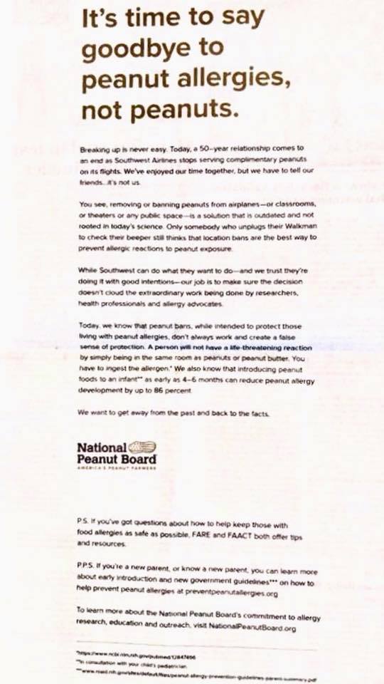
MastAttack response to the recent letter from the National Peanut Board

This post was written to directly stand against a letter recently published by the National Peanut Board (a real name that I did not make up). That letter can be viewed below and insists peanuts do not pose the risk many believe they do to allergic persons. I go through it line by line with the text from National Peanut Board prefaced with “Letter” and my thoughts prefaced with “My response.” As usual, sources are always cited.

****

Letter: It’s time to say goodbye to peanut allergies –

My response: Completely agree. Hooray! This guy for everything!

Letter: -not peanuts.

My response: Hmmm. Well, that took a jarring turn pretty quickly. That’s like saying it’s time to say goodbye to lung cancer, not cigarettes.

Letter: Breaking up is never easy. Today, a 50-year relationship comes to an end as Southwest Airlines stops serving complimentary peanuts on its flights. We’ve enjoyed our time together, but we have to tell our friends…it’s not us.

My response: Relationship drama does not belong on social media. Ten points from Slytherin.

Letter: You see, removing or banning peanuts from airplanes – or classrooms, or theaters or any public space – is a solution that is outdated and not rooted in today’s science.

My response: As a scientist, speaking on behalf of science everywhere, this is the stupidest thing I’ve ever heard. As if avoiding a potent allergen will ever be outdated. Give me a break. *rolls eyes*

Letter: Only somebody who unplugs their Walkman to check their beeper still thinks that location bans are the best way to prevent allergic reactions to peanut exposure.

My response: First of all, don’t hate. The 90’s weren’t that bad.

My response: Secondly, location bans are one piece of a complicated system that peanut allergic people utilize to find safe spaces and keep themselves out of danger. Location bans especially make sense in venues where a person can’t just leave if they find themselves confronted by a dangerous allergen. Like, I don’t know, a metal tube hurtling through the sky at warp speed?

Letter: While Southwest can do what they want to do – and we trust they’re doing it with good intentions – our job is to make sure the decision doesn’t cloud the extraordinary work being done by researchers, health professionals and allergy advocates.

My response: The extraordinary work being done is not in any way clouded by the fact that location bans are absolutely necessary in some situations. Although given the doozy that is the next paragraph, I would venture that the “problem” is that the author of this press release fundamentally misunderstands the current research findings. Hold onto your hats! This next paragraph of this press release is going to. Blow. Your. Mind. (And not in a good way)

Letter: Today, we know that peanut bans, while intended to protect those living with peanut allergies, don’t always work and create a false sense of protection.

My response: So because they don’t prevent every instance of peanut triggered anaphylaxis, we should just not have peanut bans anymore? Come on. Even the National Peanut Board has to realize how phenomenally asinine this statement is.

My response: Let’s say you are driving in a car that has four windows. Suddenly, it begins to pour. The rear passenger side window isn’t going up for some reason. Are you not going to close the other three windows to afford yourself some protection from the rain? Because this press release says they would just never even touch a window button again because when three of the windows roll up, the driver feels a false sense of security from the rain. That is how stupid this is.

My response: Anaphylaxis at school has become increasingly common.

  • “Most significant reactions in children are attributable to peanuts, fish, shellfish, egg, soy, wheat, tree nuts [and] milk.” (Sicherer 2010)
  • “Fatalities in school aged children in the United States have primarily been attributed to peanuts, tree nuts, milk, and seafood.” (Sicherer 2010)
  • “The Centers for Disease Control and Prevention recently reported an 18% increase in food allergy among school-aged children from 1997 to 2007; 1 in 25 children are now affected. Results of studies of children with food allergy indicate that 16% to 18% have experienced a reaction in school. Allergic reactions or treatment for anaphylaxis also occur in children whose allergy was previously undiagnosed (25% of cases of anaphylaxis). Fatalities were noted to be overrepresented by children with peanut, tree nut, or milk allergy.” (Sicherer 2010)
  • “In case series of fatalities from food allergy among preschool – and school-aged children in the United States., 9 of 32 fatalities occurred in school and were associated primarily with significant delays in administering epinephrine.” (Sicherer 2010)

Letter: A person will not have a life-threatening reaction by simply being in the same room as peanuts or peanut butter. You have to ingest the allergen.

My response: This is patently, verifiably false, and you should be ashamed of yourselves for saying it. You should be ashamed of OPENLY LYING about whether or not having peanuts nearby can kill someone. You should be ashamed that misinformation like this further endangers the lives of allergy patients around the world. You should be ashamed that an allergy patient might believe the words in this press release and injure themselves or worse. Where’s your false sense of security now?

My response: The American Academy of Asthma, Allergy, and Immunology (AAAAI) and the American College of Allergy, Asthma & Immunology (ACAAI) sit on a joint task force together for the purpose of developing a robust set of medical guidelines to protect patients with allergies. AAAAI and ACAAI both acknowledge that anaphylaxis can occur without ingestion of the trigger.

  • “Severe allergic reactions may be seen in some patients who only inhale or come in contact with food allergens.” (Sampson 2014)
  • “Some patients may experience symptoms on inhalation of a food allergen but not experience symptoms after ingestion of the same food allergen (eg, baker’s asthma).” (Sampson 2014)
  • “Therefore, it may be necessary to avoid food exposure by routes other than ingestion.” (Sampson 2014)
  • “In some cases, severe allergic reactions may be seen in patients who only inhale or come in contact with food allergens, thereby making avoidance even more difficult.” (Sampson 2014)
  • “Patients who are extremely allergic to peanuts might have a reaction at a ball game when peanut particles from husking are blown in the wind and inhaled by that individual and in airplanes when another passenger is eating peanuts.” (Sampson 2014)
  • “Transportation by various means also presents a risk of accidental exposure. Air travel has received the most attention, but long rail trips (especially in foreign countries) and cruise ships present their own set of risks that must be anticipated.” (Sampson 2014)

My response: Other groups also reported that anaphylaxis can occur without ingestion of the trigger.

  • “In some cases, severe allergic reactions may be seen in patients who only inhale or come in contact with food allergens, thereby making avoidance even more difficult.” (Chapman 2006)
  • “Allergic reactions that result from direct skin contact with food allergens are generally less severe than reactions due to allergen ingestion. Reactions that result from inhalation of food allergens are generally less frequent and less severe than reactions caused by either direct skin contact or ingestion. Exceptions to these generalizations are more likely in occupational environments and other settings in which food allergen sensitization occurred via either inhalation or skin contact.” (Chapman 2006) Author’s note: Please note that this was not tested on mast cell patients so reaction severity may vary.
  • “Anaphylaxis from non-ingestion exposure, such as contact with intact skin or being close to an allergen, is uncommon [but does sometimes occur].” (Lieberman 2015)
  • “Case reports and controlled studies in which foods are vaporized through heating have shown that reactions, primarily respiratory, can be elicited. These observations support limiting exposure to allergens being cooked.” (Sicherer 2010)

My response: Anaphylaxis to peanut can be life threatening.

  • “Peanut and tree nuts account for most fatal and near-fatal food allergic reactions in the United States.” (Chapman 2006)
  • “Peanut allergy affects approximately 0.6% of the general population and is the most common cause of fatal food induced anaphylaxis.” (Chapman 2006)
  • “In case series of fatalities from food allergy among preschool – and school-aged children in the United States, 9 of 32 fatalities occurred in school and were associated primarily with significant delays in administering epinephrine.” (Sicherer 2010)
  • “IgE-mediated food allergy is associated with an increased risk of death after accidental ingestion.” (Sicherer 2010)
  • “Although subsequent reactions are not necessarily more severe than initial reactions, they may be. For example, initial mild reactions to peanut may be followed by more severe reactions on subsequent exposures.” (Sicherer 2010)
  • “Clinical factors such as a history of asthma, previous reactions to trace exposures, and allergies to foods mentioned previously are potential risk factors for fatal anaphylaxis.” (Sicherer 2010)
  • “Food allergens are a frequent cause of severe anaphylaxis, particularly in patients with concomitant asthma and allergy to peanut, tree nut, or seafood. Such reactions may be biphasic or protracted. Food allergy should be considered in the differential diagnosis of patients who have idiopathic anaphylaxis.” (Sampson 2014)
  • “A study showed that peanut can be cleaned from the hands of adults by using running water and soap or commercial wipes but not antibacterial gels alone.” (Sicherer 2010)

Letter: We also know that introducing peanut foods to an infant as early as 4-6 months can reduce peanut allergy development by up to 86 percent.

My response: YOU GUYS, I FOUND THE ONLY TRUE STATEMENT ABOUT PEANUT ALLERGIES IN THIS ENTIRE PRESS RELEASE. But don’t get too excited because this only pertains to IgE mediated allergies, traditional allergies. There are a number of food allergies that are not mediated by IgE. If you have mast cell disease or eosinophilic GI disease or FPIES, the age when you introduce a food isn’t the driving issue in whether or not you tolerate it.


Sources cited

Chapman JA, et al. Food allergy: a practice parameter. Annals of Asthma, Allergy, and Immunology 2006;96:S1-S68.

Lieberman P, et al. Anaphylaxis – a practice parameter update 2015. Ann Allergy Asthma Immunol 2015;115:341-384.

Sampson HA, et al. Food allergy: A practice parameter.  J Allergy Clin Immunol  2014;134:1016-1025.

Sicherer SH, et al. Clinical Report Management of Food Allergy in the School Setting. Pediatrics 2010;126:1232-1239.


The MastAttack 107: The Layperson’s Guide to Understanding Mast Cell Diseases, Part 84

97. What is the progression of therapies to treat mast cell disease? What are the next steps if I’m not improving?

There are some general rules of thumb when adding new medications to a regimen in a mast cell patient.
Medication changes should be done one at a time. The reason for this is simple: if you get better, or worse, it will be harder to figure out which medication is the cause. Recommendations on how far apart to space changes vary, but most recommend 3-7 days.
Medications often contain dyes and inactive ingredients like lactose and alcohol that can cause reactions. Many patients take compounded medications to avoid these triggers. Bear in mind that compounding medications can be very expensive and many insurances will not cover it. You should also know that not all medications are able to be compounded.
In some instances, the likely benefit of a problem medication is enough that it is worth it to try and force tolerance. This should never be done without the supervision of a provider. Premedicating before taking a dose of the medication can help curb mast cell symptoms. Personally, I will premedicate before taking a new medication for three days. If I take it on the fourth day, when I’m not premedicated, and I react, I know that I’m unlikely to ever tolerate that med. There are also desensitization protocols that involve use of IV antihistamines, steroids, and sometimes, epinephrine.
• It is my experience that often many medications that would not typically warrant a taper down before stopping must be stopped gradually in mast cell patients to prevent reactions. For example, the general population does not need to wean off IV antihistamines. They can just stop taking it without a taper. But if a mast cell patient needed six doses of IV Benadryl, and they stop the medication cold turkey the following day, they usually react. Mast cell patients should change things slowly.
Some medications can take a while to achieve a beneficial effect. It is usually in the patient’s best interest to continue a med for long enough that they can feel confident in their assessment that a medication is or is not helping.

The following describes medication progression for overall management of mast cell disease. Please note that there are other medications that can be added for specific symptoms that are not described here. Please also note that this list is just a general guideline I find helpful.

1. Start baseline meds.

  • Start second generation H1 antihistamine, like cetirizine. Starting dose is usually one tablet 1-2 times a day.
  • Start H2 antihistamine, like famotidine. Starting dose is usually one tablet 1-2 times a day.
  • Start mast cell stabilizer, usually cromolyn. In adults, target dose is typically 200 mg four times a day. First generation H1 antihistamines, like diphenhydramine (Benadryl) are not typically used as baseline medication because they may have significant side effects, cause rebound reactions, or lose effectiveness as a rescue medication.
  • Many patients benefit from gradual dose increase in cromolyn. For reasons that are not clear, patients sometimes react to cromolyn before achieving some benefit. It can take up to four months for cromolyn to achieve full efficacy.

2. Start leukotriene blocker. Starting dose is usually one tablet at bedtime.
3. Start COX inhibitor, like aspirin. Starting dose is usually one baby aspirin (81 mg) daily. COX inhibitors interfere with production of prostaglandins.
4. Increase dose of H1 antihistamine started in step 1.
5. Increase dose of H2 antihistamine started in step 1.
6. Add another second generation H1 antihistamine to be taken along side the H1 antihistamine started in step 1. Loratadine and cetirizine are a common pair.
7. Add additional H1 antihistamine, like doxepin or cyproheptadine.
8. Start ketotifen. Starting dose is usually 1 mg twice a day. Ketotifen is both an H1 antihistamine and mast cell stabilizer. In the US, oral ketotifen must be obtained through a compounding pharmacy or imported from abroad via an FDA guidelines.
9. Increase dose of leukotriene blocker started in step 2.
10. Add first generation H1 antihistamine, hydroxyxine.
11. Start lipoxygenase inhibitor, like zileuton. Lipoxygenase inhibitors interfere with production of leukotrienes.
12. Increase dose of COX inhibitor started in step 3.
13. Increase dose of ketotifen started in step 8.
14. Take a short burst of corticosteroids, like prednisone. Corticosteroids suppress production of inflammatory mediators by mast cells.
15. Take a daily low dose of corticosteroids after a short burst at higher dose in step 14. Corticosteroids suppress production of inflammatory mediators by mast cells.
16. Start a benzodiazepine, like lorazepam.
17. Start Xolair, an anti-IgE biologic. It is unclear why Xolair helps mast cell disease when mast cell reactions typically occur without IgE involvement in this population.
18. Start regular infusion of IV fluids. This can help a lot with third spacing, fluids becoming trapped in places they aren’t supposed to, leading to swelling and functional dehydration.
19. Add therapies to block other inflammatory mast cell mediators, like interleukins or TNF. Enbrel is sometimes used in this capacity.
20. Add regular doses of IV push medications like diphenhydramine and famotidine.
21. Add continuous infusion of diphenhydramine (Benadryl) through IV line.
22. Start a tyrosine kinase inhibitor. Patients should receive regular bloodwork to monitor for organ damage or low blood cell counts.
23. Start untargeted chemo like interferon, cladribine, or hydroxyurea. Patients should receive regular bloodwork to monitor for organ damage or low blood cell counts.
24. Receive HSCT (bone marrow transplant). This option is ONLY available to patients who have malignant forms of mastocytosis and who have failed every other treatment option.

There are also a number of other changes that may help mast cell patients.
Remove obvious triggers.
Consider removing or limiting some foods. This is tricky because there are at least four different lists of low histamine foods and they all conflict. Removing lots of foods at once makes it much likelier than you will lose tolerance to them. I went strictly low histamine in 2014 and it was literally years before I could tolerate even small amounts of most of the foods I removed.
Adapt medication schedule so that triggering activities occur when medications are most available to your body. This includes things like taking antihistamines before eating or exercising.
Supplement things that many mast cell patients are deficient in. This includes vitamin D and magnesium.
• Start quercetin.
• If possible, low impact exercise can be helpful.
Manage your pain aggressively. Pain is a huge mast cell trigger.
• If you have a lot of environmental triggers, wearing a mast like a Vogmask is a good option.
• For patients who have severe GI involvement, bowel rest sometimes helps. Patients on bowel rest stop taking any food or drink by mouth and receive IV nutrition (TPN).
Avoid overtly stressful situations at all costs. I cannot emphasize this enough. Do not engage in upsetting situations if at all possible.

Sent from my iPad

Mastsisters

I love all my masto kids. But there’s something special about this little girl. The week after her second birthday, her mother, a total stranger to me, called me to convince me to be part of Addie’s team in her very complicated, very high stakes, very frightening, very literal fight for her life. I agreed. Addie was my very first case.

When our paths converged four and a half years ago, Addie was living on the edge of disaster every single day. Incidentally, so was I. We got our first PICC lines the same month. I started chemo two weeks after her mom called me. We were both in and out of the hospital with protracted anaphylaxis and scopes and procedures. We both sustained significant organ damage that year. Things were not good. But I always believed that if we could just stay alive that one day we would get better. Maybe not healthy. Maybe not well. But better. Better was the dream.

Today, we walked around Salem on a perfect breezy June day. We breathed in the salty ocean air. We looked for Halloween figurines in the small shops at Pickering Wharf. We squeezed through secret passages in dusty four hundred year old houses. We struggled to read the lettering on the smooth white faces of centuries old tombstones. We talked about school and safe foods and hospitals and ports. We ate things that would have put us in the ground when we first met.

This is not an easy life. It is never going to be easy. But there is something about looking back years later that makes you see this journey as worthwhile. We can eat food now. We aren’t admitted constantly. We are not shocking left and right. We are not always on the brink of anaphylaxis.

We made it. We are still here. We are still living in the happy moments and getting through the hard ones.

We are mastsisters. We are survivors. We are alive.

Me and Addie, Los Angeles, May 2015
Me and Addie, Salem, June 2018

Sensory memory

Sensory memory is the shortest. It lasts less than a second, usually; the memory of things you hear, echoic memory, might last a little bit longer. The things we see, hear, touch, taste, and smell all provoke a response by our nervous system. These tiny events are filtered almost immediately. Noticing everything would overwhelm our brain, both physically and figuratively. We only acknowledge things that are useful to short term memory; everything else is discarded before we even know it was there.

How many things do we see and hear and feel that our brain decides aren’t important? What if we wanted to see and hear and feel those things? What if it robbed us of these memories? I worried about this years before I understood the neurobiology of forgetting. I think that’s partly why I write: I don’t want to regret forgetting something, even if it’s not important.

My father died last month. He was diagnosed in January with a disease that would kill him. Right away, I hunted down memories of him. Hard copy photographs and pictures on our phones. Home videos from when we were growing up. Voicemails with his voice on them. Handwritten notes. Birthday cards. I started making videos of him so I could hear his voice and remember the way he moved. I journaled exhaustively, writing about my days with him in exquisite detail. I knew how easily those little pieces could be discarded and I didn’t want to lose any of it. It was too important.

All of this has had the weird side effect that I now cannot stop triggering sense memories. They just happen now. I can’t control them. I can’t choose not to pay attention. They are all so complete and so vivid. Splinters of moments that got caught in the void between the past and the present. Here but not here. Then but not then. Visible but untouchable.

In particular, I am overcome with memories from my childhood of us camping. From the ages of 4-14, my family camped every weekend from mid April to early October and for several weeks in the summer. I remember the long car rides to New Hampshire on Friday nights. Wearing my father’s blue jacket, standing in front of him around the campfire on a cool summer night. The smell of the damp earth every April on opening weekend. The sting of hitting the water after my father threw me into the air while we swam in the river. Sunny days at the end of summer when my father, my mother, my sister and I were the only people on the tiny beach. Making sand castles with moats around them. Day trips from the campground to Canobie Lake and Salisbury Beach and York Animal Kingdom.

And then the hard stuff, the memories I wish I never had to make: how soft his hair was under my hand when I tried to soothe him, the way his skin smelled at the end, how his chest rose and fell as he breathed. How it felt to wake up in a world where my father was not alive. How it was physically painful. How every day is still harder than the day before it. How it felt like I was suffocating when he died, like I’m dying. How it still feels like that.

How living in these memories is excruciating. But how losing them would be worse.