Skip to content

genetics

Genetics of MCAS: mutations and methylation

Despite having largely the same symptom profile as SM patients, people with MCAS often lack the signature D816V mutation, considered by many to be a marker of clonality and thus proliferation.  Despite the fact that their mast cells may look normal under a microscope, there is now a growing body of evidence indicating that mast cells in MCAS patients behave aberrantly due to mutations aside from D816V. 
In three studies that look at KIT mutations in MCAS patients, they were found in 26.5%, 44% and 65% of patients, respectively.  Even the average of these three values represents a significant number of people with MCAS who have KIT mutations.  Of note, these mutations are mostly outside of exon 17, where the D816V mutation is found.  In one patient, a mutation was found in the NLRP3 gene, associated with the inflammatory response. 
There are a number of other mutations in genes with various functions commonly found in patients with SM.  These include genes that make proteins to regulate other genes and genes that affect how we make proteins (epigenetic regulatory proteins, splicing machinery and transcription factors.)  To date, there have been no studies looking at whether MCAS patients have similar mutations.  However, there are clear hints that they do. 
The mutations previously mentioned were all somatic and not germline.  This means that the mutations arose after early embryonic development and thus were not heritable.  This fact indicates clearly that there are other germline mutations not yet identified that may induce the subsequent mutations.  This has been bolstered by a 2013 paper that found familial clustering in MCAS patients.   
An important finding is that nearly all mutations found in MCAS patients are heterozygous, meaning only one of the two copies was mutated.  This implies that homozygous mutations, in which both copies are mutated, could cause the cell to die.  Alternatively, the various mutations may work together to make the mutations work as strongly as if there were two mutated copies. 
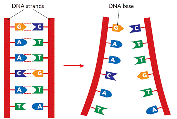
At CpG sites, the cell can add a special marker to the cytosine called a methyl group.  This is called methylation.  If the cytosine in a gene is methylated, it turns the gene off.  When a gene is turned off, your body will not use that gene or make a protein from it.  This is one type of gene regulation (epigenetics.) 
Looking at the methylation status in the genomic DNA of white blood cells from MCAS patients shows aberrant methylation patterns.  The genes incorrectly methylated included some involved in DNA/RNA repair, DNA/RNA processing, cell death, cell activity and communication with other cells.  195 individual CpG sites have been identified as candidates as markers for MCAS.  Importantly, there is a correlation between the age of symptom onset and the year of birth, which indicates a sort of anticipation of developing MCAS.  This means that gene regulation by methylation could affect acquisition of later, non-heritable mutations like the ones seen in KIT.
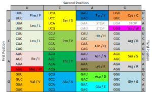
A gene is made up of introns and exons.  When an RNA code is made from a gene to tell the cell how to make a protein, the cell cuts out some pieces of that RNA.  These pieces are called introns.  The remaining pieces, which are connected back together, are called exons.  The way the cell cuts the RNA and reconnects the pieces is called splicing.   The mutations in KIT seen in MCAS patients almost all involve intron and exon junctions, where they meet.  There are also some differences in the way splicing occurs in MCAS patients. 

Reference:
Molderings, Gerhard J.  The genetic basis of mast cell activation disease – looking through a glass darkly.  2014.  Critical Reviews in Oncology/Hematology.

Gene expression and the D816V mutation

What exactly is the D816V mutation and why does it matter?  To answer that, we need to understand the basic pathway by which a cell expresses a gene.    

DNA (deoxyribonucleic acid) is the molecule that contains the genetic code for all known living organisms and some viruses.  DNA is composed of two strands that wrap around each other in a double helix pattern.  DNA is built out of nucleotides, molecules that contain energy.  The nucleotides that build DNA are adenine (A), guanine (G), thymine (T) and cytosine (C).  These nucleotides bond in specific pairs.  This means that when one nucleotide in on one strand of DNA, there is a specific nucleotide on the other strand.  A and T, and C and G specifically bond to each other.  They are known as base pairs.  DNA strands made up of base pairs are said to be “complementary.” 


RNA (ribonucleic acid) is a more versatile nucleic acid that codes, regulates and expresses genes, amongst other things.  It also has base pairs: adenine and uracil (U), and thymine and cytosine.  These nucleotides can be complementary to DNA nucleotides.  For example, an RNA adenine is complementary to a DNA thymine, and so on.

DNA replication is the process by which an exact copy of a piece of DNA is made.  This happens when a cell divides.  In replication, the DNA double helix “unzips,” or splits apart into two strands, the base pairs of which are not connected.  Special enzymes move along each of the two split strands and place the appropriate nucleotides next to each strand to form base pairs.  The end result of this is two double helices of DNA that are exact copies.   


Some parts of DNA, called genes, tell the cell how to make proteins or RNA that has a specific function.  (Sometimes RNA can also do this.)  Genes tell the cell how to build and maintain the cell and allow it pass on traits to offspring.  These proteins or RNA are made by expressing the gene.  In gene expression, the information from the gene is turned into a “gene product,” that will be made into something useful for the cell.
Transcription is the start of gene expression.  Gene expression is very complicated and controlled by many mechanisms.  Having a gene does not mean it will always be expressed.  In transcription, a piece of DNA is copied into a complementary RNA strand.   This RNA is called messenger RNA (mRNA.)  This is a complicated process with several steps.  Once a gene is translated, the mRNA with the gene code goes to the ribosome, a place in the cell that makes proteins.  Proteins are made of amino acids. 

So how exactly does the DNA code for the protein the ribosome will make? Let’s focus on that.
The ribosome reads the messenger RNA made from the DNA gene three nucleotides at a time. Again, when using the code to build a protein, the ribosome reads the code in blocks of three nucleotides. These blocks of three nucleotides are called “codons.” Every combination of three-nucleotides tells the ribosome to add a specific amino acid to the protein. The majority of genes are encoded using this same codon code. So by knowing the DNA sequence, we can anticipate the amino acids that build the protein encoded by the gene. 


How does the ribosome know where to start?  There’s a start codon.  (And some other things also.)
How does the ribosome know where to stop?  There’s a stop codon.  (And some other things also.)
There are several types of genetic mutations, or alterations of the code from the one seen in most of the population.  In a point mutation, a single nucleotide is changed.  The D816V mutation is a point mutation. 
We use a specific nomenclature to describe genetic mutations.  Amino acids are often referred to with single letter codes for the sake of brevity.  The amino acid aspartic acid is referred to as “D,” while the amino acid for valine is referred to as “V.”  In the CKIT gene, the amino acid sequence Asp-Phe-Gly (aspartic acid – phenylalanine – glycine) is very important to the receptor being shaped the right way. 
Aspartic acid is encoded by the RNA code “GAU” or “GAC.”  In cells with the D816V mutation, this sequence is changed to “GUU” or “GUC.”  The second base is changed from an A to a U.  Doing this changes the amino acid encoded from aspartic acid (D) to valine (V).  These amino acids are shaped differently, and because of this, the receptor is shaped differently and behaves differently.  When the receptor is made with the amino acid aspartic acid in that place, SCF (stem cell factor) binds to the receptor and activates the cell, telling it not to die and to make more cells.  When the receptor is made with the amino acid valine in that place, the receptor activates itself and SCF is not needed.  It basically tells itself not to die and to make more cells repeatedly. 
So the term “D816V” means that at codon 816, the code was altered in a way that changed the amino acid for aspartic acid to valine.  Some people with mast cell disease don’t have the D816V mutation, but often they have another mutation at codon 816, like D816G.  Sometimes they have a mutation somewhere else in the same “exon.”  An exon is the part of the code that is sent as RNA to be made into a gene product.  The location of the CKIT gene is referred to as exon 17.  

Image credits:


http://www.brooklyn.cuny.edu/bc/ahp/BioInfo/TT/TscriptD.html

http://genmed.yolasite.com

http://www.bristol.k12.ct.us/

http://en.wikipedia.org/wiki/Gene_expression

Mast cell disease in families

Three types of MCAD are currently known: systemic mastocytosis (SM); mast cell activation syndrome (MCAS); and mast cell leukemia (MCL).  SM and MCL are thought to be rare, while MCAS is now believed to be much more common, and possibly even the underlying cause of various clinical presentations (such as IBS and fibromyalgia.)  Very little is known about the heritability of these conditions , but many patients report that they have family members with similar symptoms. 

A study examining the familiality of MCAD found that 74% of patients interviewed had at least one first degree relative (parents, siblings, children) with systemic MCAD, regardless of MCAD subtype or gender.  The prevalence of systemic MCAD among first-degree relatives was 46%, while the prevalence in the control group is about 17%.  The prevalence of MCAD among first-degree relatives of patients with MCAS was 60%; with SM was 44%. 


MCAD subtype and severity of symptoms varied between family members.  Variable genetic alterations in CKIT were detected.  Activating CKIT mutations were found in 65% of patients, compared to 15% of the control group. The genetic mutations detected in the three families included mutations at position 816 of CKIT (D816G, D816V, S1A).  This finding is remarkable in that it disproves the longstanding belief that the somatic nature of KIT and related exon 17 mutations means that it cannot be inherited.  It also supports the belief that other mutations in genes that regulate mast cells could be contributing to these diseases.  Multiple mutations were sometimes found in the same patient, including those found in other genes (JAK2, TET2, DNMT3A, ASLX1, CBL, U2AF1, SRSF2, MS4A2). 


There was also no obvious relation between the CKIT mutations and clinical severity of MCAD.  Although familial occurrence due to shared environmental factors cannot be ruled out, it is likely that there is a significant genetic contribution to this phenomenon.  More females than males were affected.  The prevalence of MCAS was expected to be at least within the single-figure percentage range in the population (1-9%.) 


Systemic MCAD family histories include more systemic MCAD cases than would be expected when compared to the prevalence in the general population. This study advocates that the different subtypes of MCAD (MCAS and SM) should be more accurately regarded as varying types of the same disease rather than distinct diseases of mast cell dysfunction.


Reference:

Molderings GJ, Haenisch B, Bogdanow M, Fimmers R, No¨ then MM (2013) Familial Occurrence of Systemic Mast Cell Activation Disease. PLoS ONE 8(9):e76241. doi:10.1371/journal.pone.0076241