Skip to content

mast cell biology

Corticotropin releasing hormone, cortisol and mast cells

The term “HPA axis” refers collectively to the signals and feedback loops that regulate the activities of three glands: the hypothalamus, the pituitary gland, and the adrenal glands. The HPA axis is a critical component of the body’s stress response and also participates in digestion, immune modulation, emotions, sexuality and energy metabolism.

The hypothalamus is part of the brain. It performs several integral functions. It regulates metabolism, makes and releases neurohormones, and controls body temperature, hunger, thirst, circadian rhythm, sleep and energy level. It is also known to affect parenting and attachment behaviors. It effectively turns nervous system signals into endocrine signals by acting on the pituitary gland.

The pituitary gland is a small gland at the bottom of the pituitary. The anterior portion of the pituitary is part of the HPA axis. It makes and releases several hormones, including human growth hormone, thyroid stimulating hormone, adrenocorticotropic hormone (ACTH), prolactin, luteinizing hormone and follicle stimulating hormone. All of these hormones are released when hormones released by the hypothalamus act on the pituitary.

The adrenal glands are located on top of the kidneys. They primarily synthesize and release corticosteroids like cortisol and catecholamines like epinephrine and norepinephrine in response to action by the pituitary.   It also produces androgens and aldosterone.

The hypothalamus synthesizes vasopressin and corticotropin releasing hormone (CRH).   Both of those hormones stimulate the release of ACTH by the pituitary gland. ACTH stimulates the adrenals to make glucocorticoids (mostly cortisol). The cortisol then tells the hypothalamus and pituitary to suppress CRH and ACTH production. This is called a negative feedback loop.

Cortisol acts on the adrenals to make epinephrine and norepinephrine. Epi and norepi then tell the pituitary to make more ACTH, which stimulates the production of cortisol.

When you take steroids regularly, it suppresses ACTH so that your body stops making its own steroids. This is why weaning steroids is very important. By weaning, your body should gradually start making its own cortisol to replace the deficit when you lower your steroid dose. However, this doesn’t always work. People who do not make enough cortisol on their own are called adrenally insufficient and are steroid dependent. People with this condition can suffer “Addisonian crises” if their steroid levels drop dangerously low. This is a medical emergency.

CRH is released by the hypothalamus in response to stress. This drives the production of cortisol to help manage stressful situations of either a physical or emotional nature. Mast cell attacks and anaphylaxis are examples of physically stressful situations that stimulate release of CRH.

CRH binds to CRHR-1 and CHRH-2 receptors on various cells, including mast cells. When it binds to mast cells, it stimulates the release of VEGF, but not histamine, tryptase or IL-8. This type of release is called selective release as it does not involve the release of preformed granules (degranulation.) Additionally, CRH is also released by mast cells. This can act on the mast cells or other cells with CRHR receptors, like those in the pituitary. The exact purpose of mast cells releasing CRH is not clear.


References:

Theoharis C. Theoharides, et al. Mast cells and inflammation. Biochimica et Biophysica Acta 1822 (2012) 21–33.
 

Lesser known mast cell mediators (Part 4)

Interleukin-1a (IL-1a) is largely responsible for inflammation, fever and sepsis. It activates TNF-a and the work very closely together. Their cofunctions include PGE2 synthesis, nitric oxide production, insulin resistance and IL-8 and chemokine production.

Interleukin-1b (IL-1b) has been implicated in several autoinflammatory syndromes. It is also important in cell proliferation, differentiation and apoptosis. Its induction of COX2 cytokine in the nervous system contributes to inflammatory pain hypersensitivity.

Interleukin-2 (IL-2) is crucial in prevention of autoimmune disease by regulating T cell differentiation. It is also thought to be involved in itchiness and psoriasis. IL-2 is used in the treatment of cancers.

Interleukin 3 (IL-3) drives the differentiation of multipotent hematopoietic stem cells into myeloid progenitor cells. If IL-7 is also present, they can work synergistically to trigger differentiation into lymphoid progenitor cells. IL-3 induces proliferation of all myeloid cells (including mast cells) along with other cytokines like IL-6. It supports growth and differentiation of T cells from bone marrow when an immune response is triggered.

Interleukin 4 (IL-4) changes naïve T cells to T helper cells, which secrete chemicals to drive actions of other immune cells. T helper cells then secrete additional IL-4 to perpetuate the cycle. IL-4 participates in the airway inflammation seen in allergic asthma.

Interleukin 5 (IL-5) encourages growth of B cells and antibody secretion as well as eosinophil activation. It is heavily involved in allergic diseases, particularly those in which eosinophils are notably increased. Mepolizumab is a monoclonal antibody against IL-5 that can reduce excessive eosinophils.

Interleukin 6 (IL-6) mediates fever and the acute phase inflammatory response. It is secreted to stimulate bone resorption and inhibitors of IL-6 are used to treat osteoporosis (including estrogen.) It inhibits TNF-a and IL-1. Unusually, it also has anti-inflammatory behaviors, particularly during exercise in the muscle.

Interleukin 9 (IL-9) increases cell proliferation and impedes apoptosis, cell death, of hematopoietic cells. It is particularly important in asthma and bronchial hyperresponsiveness.

Interleukin 10 (IL-10) is an anti-inflammatory molecule involved in regulating the JAK-STAT pathway. It counteracts many of the inflammatory effects of mast cells, often by interfering with production of substances like interferons and TNF-a.   Exercise increases levels of this molecule.

Interleukin 13 (IL-13) is critical in initiation of airway disease. It induces matrix metalloproteinases to act. IL-13 can also induce IgE release from B cells. It is effectively a link between allergic inflammatory cells and the non-immune cells they interact with. Excessive , IL-13 causes airway hyperresponsiveness, goblet cell metaplasia and oversecretion of mucus.


Lesser known mast cell mediators (Part 3)

Substance P is a neurotransmitter and modulates neurologic responses. It is found in many sensory nerves as well as the brain and spinal cord. It participates in inflammatory responses and is important in pain perception. It is involved in mood disorders, anxiety, stress, nerve growth, respiration, neurotoxicity, nausea, vomiting and pain perception. Its release from nerve fibers into the skin, muscle and joints is thought to cause neurogenic inflammation.

Urocortin is related to corticotropin releasing factor (CRF.) It strongly suppresses blood pressure and increases coronary blood flow. It is thought to have a role in increasing appetite during times of stress.

VEGF-A (vascular endothelial growth factor A) is a member of the platelet derived growth factor (PDGF)/VEGF family. It is important in nerve biology and is the substance mainly involved in inducing growth of blood vessels. It is heavily involved in diseases that involve blood vessels, like diabetic retinopathy and macular degeneration. It is a vasodilator and increases permeability of the smaller vessels.

VIP (vasoactive intestinal peptide) is a small protein like molecule used by nerve cells for communication. It stimulates heart contraction, vasodilation, lowers blood pressure, and relaxes the smooth muscles of the trachea, stomach and gall bladder. It also inhibits gastric acid secretion and absorption in the intestine.

Mast cell kininogenase removes a portion of a compound to release active bradykinin. This is important in the kinin system.

Phospholipase A2 promotes inflammation by initiating formation of arachidonic acid, the precursor needed to form many inflammatory molecules, including prostaglandins. Excessive levels of phospholipase A2 can lead to increased vascular inflammation, such as a seen in coronary artery disease and acute coronary syndrome. Elevated PLA2 is found in the cerebrospinal fluid of people with Alzheimer’s disease and multiple sclerosis.

Corticotropin releasing hormone (CRH) is a hormone and neurotransmitter. High CRH levels have been associated with Alzheimer’s disease and severe depression. CRH is produced in the hypothalamus and is carried to the pituitary gland, where it stimulates secretion of adrenocorticotropic hormone (ACTH.) ACTH drives synthesis of cortisol and other steroids. Imbalance of these hormones can have dire consequences.

Endothelin is the most potent vasoconstrictor currently described. It raises blood pressure and if uncontrolled, hypertension may result. It is involved in many disease processes, including cardiac hypertrophy, type II diabetes and Hirschsprung disease.

Chondroitin is found largely in connective tissues and is a principal component of cartilage. It is typically bound to other components when released from mast cells and interacts with a variety of molecules.

Hyaluronic acid is widely found in epithelial, neural and connective tissues. It participates in a variety of reactions and sees significant turnover daily. When hyaluronic acid is degraded as part of the turnover, its degradation products can cause inflammatory responses.

Mast cells, heparin and bradykinin: The effects of mast cells on the kinin-kallikrein system

The kinin-kallikrein system is a hormonal system with effects on inflammation, blood pressure, coagulation and pain perception. This system is known to have a significant role on the cardiovascular system, including cardiac failure, ischemia and left ventricular hypertrophy. Despite significant research, it is not entirely understood.

Kininogens are proteins that have extra pieces on them. Kininogenases cut off those extra pieces. Active kinins that can act on the body are the result of this action. So kininogenases change kininogens to form kinins.

There are two types of kininogens: low molecular weight (smaller) and high molecular weight (larger.) We are going to focus on HMW, which circulates in the blood.

Also circulating in the blood are two other components called prekallikrein (sometimes called Fletcher factor) and Hageman factor (Factor XII.) When Hageman factor lands on a negatively charged surface, it changes shape and becomes Factor XIIa. Factor XIIa changes the prekallikrein to kallikrein. Kallikrein is a kininogenase.

When kallikrein finds a kininogen, it cuts off the extra piece to release bradykinin. Bradykinin is a kinin and is ready to act on the body.

Bradykinin has several functions in the body. It contributes to contractility of duodenum, ileum and cecum. In the lungs, it can cause chloride secretion and bronchoconstriction. It can cause smooth muscle contraction in the uterus, bladder and vas deferens. It contributes to rheumatoid arthritis, inflammation, pain sensation and hyperalgesia. It also induces cell proliferation, collagen synthesis, and release of nitric oxide, prostacyclin, TNF-a and interleukins. It can also cause release of glutamate by nerve cells. Glutamate has a variety of actions in the body and excessive release can cause epileptic seizures, ALS, lathyrism, autism and stroke.

Bradykinin acts on the endothelium, the cells that line the inner surface of blood and lymphatic vessels, to cause the blood vessels to dilate. This decreases blood pressure. It also regulates sodium excretion from the kidneys, which can further decrease blood pressure. Kininogen levels are reduced in hypertensive patients. Kinins, including bradykinin, oppose the action of angiotensin II, a hypertensive agent.

So how are mast cells related to this system? A couple of ways. The first way is that they release kininogenases and bradykinin. Tryptase can actually behave as a kininogenase. The second way is by being the exclusive producers of heparin.

As I mentioned above, Factor XII needs to change to Factor XIIa to initiate the formation of bradykinin. It does this when it contacts a negatively charged surface. In the lab, you can use a surface like glass for this. But in the body, it often happens on the surfaces of large, negatively charged proteins like heparin. (Side note: Factor XII is part of the clotting cascade. It can be activated by medical devices like PICC lines and that is why they carry a risk of clot formation.) So by releasing heparin, mast cells cause the formation of bradykinin. When the mast cells release heparin in inappropriate amounts, too much bradykinin is formed.

Overproduction of bradykinin is one of the principal causes of angioedema. In hereditary angioedema, the body is deficient in a component that regulates bradykinin. One of the reasons that physical trauma can cause mast cell degranulation is because it causes formation of bradykinin. Bradykinin in turn causes mast cell degranulation with release of histamine and serotonin, among other contents.

Bradykinin antagonists are being researched as possible therapies for hereditary angioedema. Icatibant is one such medication. Bromelain, found in the stems and leaves of pineapples, are known to suppress swelling caused by bradykinin. Aloe and polyphenols, like those in green tea, are also known to suppress bradykinin activity.

References:

Kaplan AP, Ghebrehiwet B. The plasma bradykinin-forming pathways and its interrelationships with complement. Mol Immunol. 2010 Aug; 47(13):2161-9

Oschatz C, et al. Mast cells increase vascular permeability by heparin-initiated bradykinin formation in vivo. Immunity. 2011 Feb 25; 34(2):258-68.


Brunnée T, et al. Mast cell derived heparin activates the contact system: a link to kinin generation in allergic reactions. Clin Exp Allergy. 1997 Jun;27(6):653-63.


Lesser known mast cell mediators (Part 2)

Arylsulfatase A, also called cerebroside sulfatase, breaks down compounds to yield cerebrosides and sulfates. Cerebrosides can be either galactocerebrosides, which are found in all tissues of the nervous system; or glucocerebrosides, which are found in the skin, spleen, red blood cells and, to a lesser extent, tissues of the nervous system.

Arylsulfatase B, which has several other names, breaks down large sugar compounds, especially dermatan sulfate and chondroitin sulfate. Arylsulfatase B is mostly found in the liver, pancreas and kidneys.

Mutations in the gene for either arylsulfatase can lead to a variety of heritable disorders, including mucopolysaccharidosis VI and metachromatic leukodystrophy.

Chymases include mast cell protease 1, mast cell serine proteinase, skeletal muscle protease and so on. They are found almost exclusively in mast cells, but are present in small amounts in the granules of basophils. They have several functions, including generating an inflammatory response to parasites. They convert angiotension I to angiotensin II and therefore impact hypertension and atherosclerosis.

Bradykinin causes dilation of blood vessels, which induces a corresponding drop in blood pressure. It achieves its action by triggering release of prostacyclin, nitric oxide and endothelium derived hyperpolarizing factor. It also causes contraction of non-vascular smooth muscles in the respiratory and GI tracts, and is involved in the way the body senses pain. Bradykinin is important in angioedema.

Angiogenin, also called ribonuclease 5, stimulates the formation of new blood vessels. It drives the degradation of the basement membrane and local matrix so that endothelial cells can move toward the vascular spaces.

Leptin is the hormone that regulates hunger. It is mostly produced by fat cells, but is released by mast cells as well. When a specific amount of fat is stored in the body, leptin is secreted and tells the brain that it is full. It opposes the action of ghrelin, the hormone that tells your body it is hungry.

Renin, also called angiotensinogenase, is a critical component of the renin-angiotension system (RAS) that controls the volume of fluids not in cells, including blood plasma, lymph and interstitial fluid. It regulates the body’s mean arterial blood pressure. It converts angiotensinogen to angiotensin I.

Somatostatin, also growth hormone inhibiting hormone (GHIH), regulates the endocrine system, transmission of neurologic signals and cell growth by acting on somatostatin receptors and inhibiting the release of various secondary hormones. It inhibits secretion of glucagon and insulin. It is secreted throughout the GI system and decreases stomach acid production by downregulating the release of gastrin, secretin and histamine.

Lesser known mast cell mediators (Part 1)

I have posted at length about the roles of histamine and serotonin. Here are some less well known mast cell mediators. I will be doing in depth posts on the more relevant substances in the near future.

Monocyte chemotactic protein 1 (MCP-1), also known as chemokine ligand 2 (CCL2), draws other white blood cells, including memory T cells, monocytes and dendritic cells, to the site of injury or infection. It has important functions in neuroinflammation as seen in experimental autoimmune encephalitis, traumatic brain injuries, epilepsy and Alzheimer’s disease; and in diseases with pathologic infiltration of monocytes, like rheumatoid arthritis.

Chemokine ligand 3 (CCP7) recruits monocytes and regulates macrophage activity. It is known to interact with MMP2.

MMP2 (matrix metalloproteinase 2) is involved in tissue remodeling, reproduction and fetal development. It degrades type IV collagen. It has regulatory effects on the menstrual cycle and has been tied to growth of new blood vessels.

Interleukin 8 (IL-8), also known as neutrophil chemotactic factor (NCF), draws other white cells, mostly neutrophils, to a site of infection. It can activate multiple cells types, including mast cells, and promotes degranulation. It has been linked to bronchiolitis, psoriasis and inflammation.

MCP-4 (CCL13) attracts T lymphocytes, eosinophils, monocytes and basophils to an area of inflammation. Improper regulation can exacerbate asthma symptoms. Mast cells can release MCP-1 when stimulated by TNF-a and IL-1.

CCL5 (RANTES) attracts T cells, eosinophils and basophils. When IL-2 and interferon-γ are present, CCL5 activates natural killer cells and causes proliferation of the same. It is also important in bone metabolism.

CCL11 (eotaxin-1) specifically recruits eosinophils and is heavily involved in allergic inflammatory responses.

CPA3 (carboxypeptidase A3) digests proteins. It is released complexed with heparin proteoglycan along with chymase and tryptase.

Both interferon α (IFN- α) and interferon β (IFN-β) are made in response to viral infections. Their activities are regulated by IFN- γ. IFN- γ also draws white cells to the site of inflammation. Failure to properly regulate interferon levels can cause autoimmune disease. Interferons are so called because of their ability to “interfere” with viral infection. They are responsible for “flu type symptoms,” such as fever, muscle aches and lethargy.

All mediators listed here are produced by mast cells and stored in granules until degranulation.


Mast cells and cardiac and vascular dysfunction

Mast cells have been implicated in several types of cardiac and vascular dysfunction.  Mast cells are thought to contribute to rupture of atherosclerotic plaques by mediator release.  They are found around blood clots in the body.  Mast cells may destabilize them and mature them by releasing heparin and degrading fibrinogen with tryptase. Increased numbers of mast cells are associated with coronary vasospasm. 
Mast cell mediator levels are often higher in vascular and cardiac events.  In patients who die from coronary heart disease, the histamine concentration in the coronary artery than in control subjects.  Higher white blood cell, platelet and plasma histamine levels are found in patients with peripheral vascular disease. Increased histamine levels are found in patients with both stable coronary artery disease and acute coronary syndrome.  Plasma histamine is elevated in the great cardiac vein of 8/11 patients with variant angina. 
One study found that tryptase is higher in patients without acute coronary syndrome undergoing catheterization, compared to patients with and without obstructive coronary disease.  In this study, patients in the highest 25% of tryptase values had 4.3x greater risk for coronary artery disease.  Tryptase is being investigated as a marker to identify asymptomatic patients with coronary artery disease and to track efficacy of treatment. 
Mast cell mediators are also elevated in non-allergic coronary events, indicating that there is a common pathway for both allergic (Kounis syndrome) and non-allergic cardiac episodes.  Two cholesterol lowering medications, cervistatin and atorvastatin, inhibit stem cell factor (SCF) mediated differentiation of mast cells.  Lovastatin inhibited IgE-mediated degranulation.

References:
Ribatti D, Crivellato E. Mast cells, angiogenesis, and tumour growth. Biochim. Biophys. Acta Mol. Basis Dis. 2012 Jan; 1822(1):2-8.
Glowacki J, Mulliken JB. Mast cells in hemangioma and vascular malformations. Pediatrics 1982; 70(1):48-51.
Kolck UW, Alfter K, Homann J, von Kügelgen I, Molderings GJ. Cardiac mast cells: implications for heart failure. JACC 2007 Mar 13; 49(10):1106-1108.
Biteker M.  Current understanding of Kounis Syndrome.  Expert Rev Clin Immunol 2010 Sep;6(5):777-88.

Gene expression and the D816V mutation

What exactly is the D816V mutation and why does it matter?  To answer that, we need to understand the basic pathway by which a cell expresses a gene.    

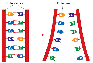
DNA (deoxyribonucleic acid) is the molecule that contains the genetic code for all known living organisms and some viruses.  DNA is composed of two strands that wrap around each other in a double helix pattern.  DNA is built out of nucleotides, molecules that contain energy.  The nucleotides that build DNA are adenine (A), guanine (G), thymine (T) and cytosine (C).  These nucleotides bond in specific pairs.  This means that when one nucleotide in on one strand of DNA, there is a specific nucleotide on the other strand.  A and T, and C and G specifically bond to each other.  They are known as base pairs.  DNA strands made up of base pairs are said to be “complementary.” 


RNA (ribonucleic acid) is a more versatile nucleic acid that codes, regulates and expresses genes, amongst other things.  It also has base pairs: adenine and uracil (U), and thymine and cytosine.  These nucleotides can be complementary to DNA nucleotides.  For example, an RNA adenine is complementary to a DNA thymine, and so on.

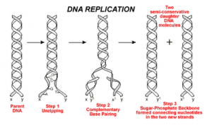
DNA replication is the process by which an exact copy of a piece of DNA is made.  This happens when a cell divides.  In replication, the DNA double helix “unzips,” or splits apart into two strands, the base pairs of which are not connected.  Special enzymes move along each of the two split strands and place the appropriate nucleotides next to each strand to form base pairs.  The end result of this is two double helices of DNA that are exact copies.   


Some parts of DNA, called genes, tell the cell how to make proteins or RNA that has a specific function.  (Sometimes RNA can also do this.)  Genes tell the cell how to build and maintain the cell and allow it pass on traits to offspring.  These proteins or RNA are made by expressing the gene.  In gene expression, the information from the gene is turned into a “gene product,” that will be made into something useful for the cell.
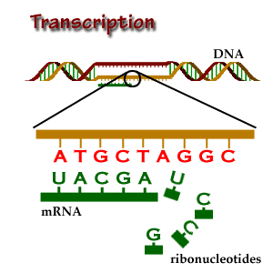
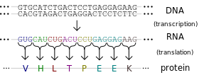
Transcription is the start of gene expression.  Gene expression is very complicated and controlled by many mechanisms.  Having a gene does not mean it will always be expressed.  In transcription, a piece of DNA is copied into a complementary RNA strand.   This RNA is called messenger RNA (mRNA.)  This is a complicated process with several steps.  Once a gene is translated, the mRNA with the gene code goes to the ribosome, a place in the cell that makes proteins.  Proteins are made of amino acids. 

So how exactly does the DNA code for the protein the ribosome will make? Let’s focus on that.
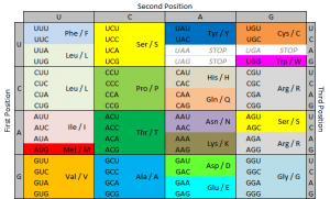
The ribosome reads the messenger RNA made from the DNA gene three nucleotides at a time. Again, when using the code to build a protein, the ribosome reads the code in blocks of three nucleotides. These blocks of three nucleotides are called “codons.” Every combination of three-nucleotides tells the ribosome to add a specific amino acid to the protein. The majority of genes are encoded using this same codon code. So by knowing the DNA sequence, we can anticipate the amino acids that build the protein encoded by the gene. 


How does the ribosome know where to start?  There’s a start codon.  (And some other things also.)
How does the ribosome know where to stop?  There’s a stop codon.  (And some other things also.)
There are several types of genetic mutations, or alterations of the code from the one seen in most of the population.  In a point mutation, a single nucleotide is changed.  The D816V mutation is a point mutation. 
We use a specific nomenclature to describe genetic mutations.  Amino acids are often referred to with single letter codes for the sake of brevity.  The amino acid aspartic acid is referred to as “D,” while the amino acid for valine is referred to as “V.”  In the CKIT gene, the amino acid sequence Asp-Phe-Gly (aspartic acid – phenylalanine – glycine) is very important to the receptor being shaped the right way. 
Aspartic acid is encoded by the RNA code “GAU” or “GAC.”  In cells with the D816V mutation, this sequence is changed to “GUU” or “GUC.”  The second base is changed from an A to a U.  Doing this changes the amino acid encoded from aspartic acid (D) to valine (V).  These amino acids are shaped differently, and because of this, the receptor is shaped differently and behaves differently.  When the receptor is made with the amino acid aspartic acid in that place, SCF (stem cell factor) binds to the receptor and activates the cell, telling it not to die and to make more cells.  When the receptor is made with the amino acid valine in that place, the receptor activates itself and SCF is not needed.  It basically tells itself not to die and to make more cells repeatedly. 
So the term “D816V” means that at codon 816, the code was altered in a way that changed the amino acid for aspartic acid to valine.  Some people with mast cell disease don’t have the D816V mutation, but often they have another mutation at codon 816, like D816G.  Sometimes they have a mutation somewhere else in the same “exon.”  An exon is the part of the code that is sent as RNA to be made into a gene product.  The location of the CKIT gene is referred to as exon 17.  

Image credits:


http://www.brooklyn.cuny.edu/bc/ahp/BioInfo/TT/TscriptD.html

http://genmed.yolasite.com

http://www.bristol.k12.ct.us/

http://en.wikipedia.org/wiki/Gene_expression

Mast cells, eosinophils and the perfect storm of inflammation

Mast cells and eosinophils have a lot of common functions.  In allergic and inflammatory states, these cells come into physical contact with each other, as well as communicate using chemical signals called cytokines and chemokines.  Mast cells and eosinophils are often found together in affected tissues in disorders like allergic rhinitis, atopic dermatitis, and asthma.  Mast cells initiate the allergic inflammatory response once activated.  This signals for eosinophils to come to the tissue.  Increased numbers of mast cells and eosinophils are found in diseases like eosinophilic esophagitis, chronic gastritis, GI neoplasms, parasitic infections and IBD.  Both mast cells and eosinophils respond to eotaxins, molecules that draw eosinophils to the inflamed area.  So one signal causes both cell types to go to the affected tissue. 

Mast cells and eosinophils interact a lot by using chemicals.  Mast cell released heparin stabilizes eotaxins.  Mast cells produce IL-3 and IL-5, which lengthen the lives of eosinophils in tissue.  Mast cell mediator chymase suppresses eosinophil death and causes eosinophils to release several chemicals.   Tryptase can limit eosinophil activation.  In turn, eosinophils produce stem cell factor (SCF), which attract mast cells and protects them from cell death.  Both cell types express some common receptors, like Siglec-8, which induces eosinophil death and inhibits IgE-mediated mast cell activation.  Interactions between these cells increase activation and proliferation. 
Patients with SM may have another blood disorder, including CEL or hypereosinophilic syndrome (HES.)  SM-HES and SM-CEL with the D816V CKIT mutation has been found, and the mutation is present in both the mast cells and the eosinophils.  However, it is likely that the FIP1L1-PDGFRA fusion gene (an aberrant tyrosine kinase) is the cause of the coexistent eosinophilic and abnormal mast cell proliferations.  The FIP1L1-PDGFRA fusion has been found in several cell types, including neutrophils, monocytes and mast cells.  This finding is consistent with a mutational origin in a blood stem cell that makes mutated mast cells and overproduces eosinophils.  When these cells are not neoplastic, they are derived from separate stem cell lineages.
Shortly after the discovery of this fusion gene, there was significant debate over whether FIP1L1-PDGFRA+ disease was an eosinophilic neoplasm with increased mast cells or systemic mastocytosis with eosinophilia.  Patients with FIP1L1-PDGFRA+ eosinophilia have a lot of symptoms in common with SM: swollen spleen, hypercellular bone marrow, high numbers of abnormally shaped bone marrow cells, marrow fibrosis and elevated serum tryptase.  However, these bone marrows show less dense clusters of mast cells.  In some cases, mast cells were spindled and expressed CD2 or CD25.  Still, the WHO considers it a distinct entity and not a subset of SM.
In CKIT+ patients, GI symptoms, UP, thrombocytosis, serum tryptase value, and dense mast cell clusters aggregates in bone marrow are significantly increased.  Cardiac and pulmonary symptoms, eosinophilia, eosinophil to tryptase ratio, elevated serum B12 and male sex were higher in FIP1L1-PDGFRA+ group.
Eosinophilia in SM patients has no effect on prognosis.  Eosinophilia in MDS patients predicted significantly reduced survival.  In T lymphoblastic leukemia, eosinophilia was unfavorable for survival.  Density and activation of tissue eosinophils is related to disease progression in several neoplasms.  Mast cells and eosinophils are found in increased numbers in neoplastic disorders like Hodgkin lymphoma. 
Presence of FIP1L1-PGDFRA indicates treatment with imatinib (Gleevec), regardless of organ dysfunction.  It can show remission within 4 weeks, even at low doses.  Some patients with CKIT+ SM with HES or CEL have rapid and complete normalization of severe eosinophilia with midostaurin treatment. 

Reference:
Gotlib, Jason, Akin, Cem.  2012.  Mast cells and eosinophils in mastocytosis, chronic eosinophilic leukemia, and non-clonal disorders.  Semin Hematol 49:128-137. 

Effect of anemia on mast cells

A paper released in September 2012 addressed the effect of iron availability on mast cell degranulation.
Inside the bodies of mice, it was observed that mice with decreased iron stores had more severe inflammatory reactions.  Importantly, iron supplementation decreased the severity of the inflammation, particularly in the context of allergic asthma.  Increased iron inhibited the production of inflammatory molecules in pulmonary tissues, including various interleukins and interferons. 
Outside of the body, mast cells were incubated with and without iron for 30 minutes.  IgE was then added to activate the mast cells.  The mast cells that were incubated with iron degranulated 30% less than those without iron present.  Spontaneous degranulation, without IgE crosslinking, was not affected.  The presence of iron also dramatically affected the production of inflammatory molecules by mast cells.  Production of TNF, MCP-1 and IL-6 decreased by 94%, 29% and 27%, respectively.  MCP-1 attracts macrophages. 
Iron supplementation decreased the severity of allergic asthma, and decreased mast cell degranulation by IgE crosslinking 30%, as well as decreasing production of inflammatory molecules by mast cells.

Reference:
Hale LP, Kant EP, Greer PK, Foster WM (2012) Iron Supplementation Decreases Severity of Allergic Inflammation in Murine Lung. PLoS ONE 7(9): e45667. doi:10.1371/journal.pone.0045667