Skip to content

epigenetics

Mast cell mutations: TET2 and mutation profiles of aggressive subtypes

TET2 (Tet methylcytosine dioxygenase 2) is found to be mutated in 20.8-29% of SM patients. Of note, dozens of mutations have been identified in this gene, including missense, nonsense, frameshift and deletion mutations. These mutations cause formation of a defective and less active TET2 enzyme. TET2 is located at chromosome 4q24 and mutations at this location are associated in both MPN and MDS conditions.

TET2 is involved in DNA methylation and demethylation, although the exact nature of this involvement is not clear. When a methyl group is added to a cytosine at a specific place in front of a gene, the gene is turned off and is not expressed. This is called “methylation.” TET2 adds a hydroxyl group to 5-methylcytosine, but it is not well understood if this turns the gene off. TET2 may also be involved in demethylating DNA, or removing those specific methyl groups. It has been shown to be involved with DNA demethylation during bone development.

One study looked at the mutational profiles of patients with various forms of SM, including ISM, SSM, SM-AHNMD, ASM and MCL, all of whom were positive for CKIT D816V mutation. 15/39 had a TET2 mutation. None of those patients had ISM or SSM. Of those with an aggressive form and a TET2 mutation, 67% had more than one TET2 mutation.

In this study, 24/27 patients with advanced SM (SM-AHNMD, ASM, MCL) had mutations beyond the D816V mutation. 5/5 SM-AHNMD patients and 19/22 ASM or MCL patients had multiple mutations (CKIT and something else.) In contrast, only 3/12 ISM or SSM patients had additional mutations. In advanced SM, 78% had at least 3 mutations, and 41% had at least 5.

These mutational profiles have clear implications clinically. 96% patients with major blood abnormalities (anemia <10 g/dL and/or thrombocytopenia < 100 x 10e9/L in addition to monocytosis > 1 x 10e9/L and/or eosinophilia >10%) had at least one additional molecular mutation regardless of SM subtype.

Advanced SM patients in this study all had one of the following multiple mutation profiles: 26% KIT-TET2-SRSF2, 18% KIT-SRSF2-RUNX1, 13% KIT-TET2-CBL, 10% KIT-SRSF2-ASXL1 10%, and 10% KIT-TET2-ASXL1. Patients with advanced SM (and therefore multiple mutations) were also found to be significantly older (68 years of age on average) than those with just the CKIT mutation (48 years of age on average.)

Having a TET2 mutation seems to predispose myeloid cells to become neoplastic later in life. It is important to distinguish that the TET2 mutation seems to “allow” this transformation rather than causing it. In mice who don’t have the TET2 gene and thus don’t have the TET2 enzyme, stem and progenitor cells have trouble maintaining balance and spontaneously become neoplastic later in life. In TET2 deficient cells, mast cells with D816V mutation show increase in proliferation and survival as opposed to those without with normal TET2 levels. Presence of TET2 in addition to the presence of CKIT D816V mutation is associated with more aggressive forms of SM (including ASM, MCL and SM-AHNMD.)


References:

Damaj, G., Joris, M., Chandersris, O., Hanssens, K., Soucie, E., Canioni, D., et al., 2014.ASXL1 but not TET2 Mutations Adversely Impact Overall Survival of PatientsSuffering Systemic Mastocytosis with Associated Clonal Hematologic Non-Mast-Cell Diseases. PLoS ONE 9 (1), e85362.

Schwaab, J., Schnittger, S., Sotlar, K., Walz, C., Fabarius, A., Pfirrmann, M., et al., 2013.Comprehensive mutational profiling in advanced systemic mastocytosis. Blood122 (October (14)), 2460–2466.

Soucie, E., Hanssens, K., Mercher, T., Georgin-Lavialle, S., Damaj, G., Livideanu, C.,et al., 2012. In aggressive forms of mastocytosis. TET2 loss cooperates with c-KITD816V to transform mast cells. Blood 120 (December (24)), 4846–4849.

Soucie, E., Brenet, F., Dubreuil, P. Molecular basis of mast cell disease. Molecular Immunology 63 (2015) 55-60.

Food allergy series: Risk factors for developing food allergies

There are a number of factors that seem to contribute to developing food allergies. Genetics seems to play an important role. One study found that that 64% of monozygotic twins had a concordance of peanut allergy, while only 7% dizygotic twins concorded. This means that in 64% of identical twin sets, either both had peanut allergies or neither did, while that was the case in only 7% of fraternal twins. Because monozygotic twins have identical genetic sequences, this finding implies a strong genetic component. HLA haplotyping has been studied, with conflicting reports on links between HLA type and allergies.

One of the most important genetic findings regards filaggrin, a skin barrier protein. Patients with a specific filaggrin mutation are more likely to develop peanut sensitization. This indicates that a damaged skin barrier could cause food sensitization and allergy, and further supports the idea that non-oral exposures can be sensitizing. Additionally, filaggrin mutation causes increased inflammatory mediators in the skin.

Generally, children with peanut or tree nut allergies react the first time the food is ingested. It is thought that they previously encountered these allergens in their environment. Household exposure to peanut was a significant risk factor for peanut allergy in infants. Peanut responsive T cells are found in the skin homing T cells in peanut allergic patients, implying that patients may first be exposed through the skin. There is not yet enough data on maternal ingestion of allergens to know if this is a risk factor. There are conflicting data sets on whether breastfeeding is protective against food allergies, and in any case, outcome appears to be dependent on the mother’s own sensitivity profile. Now seen in multiple recent studies, it seems that early oral exposure to food allergens may actually be protective against food allergy, a change from data produced over a decade ago.

Immune dysregulation is obviously involved in food allergies. Low vitamin A and vitamin D, which modulate the immune system, have been noted as risk factors. Interestingly, food allergy frequency varies with latitude, indicating a further possible connection to sun exposure and vitamin D deficiency. High fat diet can also change the composition and behavior of the microbial content of the GI microbiome. Medium chain triglycerides can increase sensitization when given along with food antigens in mice. There are mixed results with long chain fatty acids.

The changes in hygiene, cleaning products and use of antimicrobial compounds by the general public in the last decades have been implicated in many of the immune changes we have seen, including increasing autoimmune diseases and food allergies. This is known as the hygiene hypothesis, and it states that reduced exposure to microbes changes immune defense, causing improper reactivity to harmless components, like food and self cells. In food allergic mice, the gut microbiota has a very specific composition and transferring this flora set can actually make others more likely to develop food allergies. Dysbiosis has been noted in children with food allergies and a sequencing study demonstrated that food allergic children with atopic dermatitis have reduced microbial diversity in the gut.


References:

Cecilia Berin, Hugh A. Sampson. Food allergy: an enigmatic epidemic. Trends in Immunology, Volume 34, Issue 8, August 2013, pages 390-397.

Cecilia Berin, Hugh A. Sampson. Mucosal Immunology of Food Allergy. Current Biology, Volume 23, Issue 9, May 2013, pages R389-R400.

MTHFR, folate metabolism and methylation

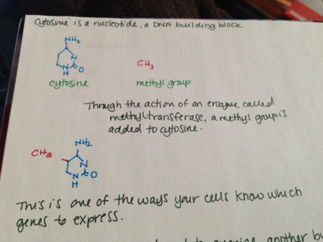
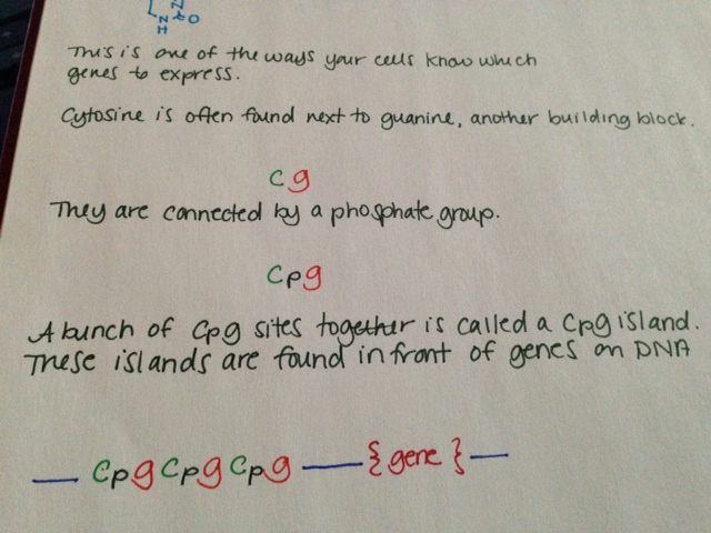
MTHFR (methylenetetrahydrofolate reductase) is an enzyme involved in folate metabolism. It is rate limiting, which means that if there is not enough, your body cannot metabolize enough folate; if there is too much, it metabolizes too much. How much folate your body is able to metabolize is directly related to how much MTHFR you have in your body.   Some folate broken down by your body is used to methylate DNA. There has been much conflicting evidence, but it seems that low folate in the body is associated with less DNA methylation overall, which may be associated with cancer (Crider, 2012.)

A single nucleotide polymorphism (SNP) is a change in DNA sequence in which one nucleotide (a DNA building block) is changed. Importantly, SNPs are common. In fact, they are so common that the way your body codes its DNA allows for SNPs to not change gene expression in many cases. This is called wobble. Three DNA building blocks in a row make one amino acid, which is used to build proteins that do things in the body. However, in many cases, the third building block can be any building block and it will still make the same amino acid. Please consider this. SNPs are so common that your cells know that in many cases, 1 in every 3 DNA building blocks can be anything and it won’t change a thing.

SNPs have become the topic of increased interest, both by medical and scientific professions and by lay people. It is certainly true that some SNPs play a role in development and progression of diseases. MTHFR has been found to have up to 24 reported SNPs, with two being of particular interest. These are C677T and A1298C.

The normal (or “wildtype”) form of MTHFR has a cytosine (C) nucleotide where people with the C677T mutation have a thymine nucleotide. If you have two copies of the regular gene, you are 677CC and homozygous for the regular form of the gene (the allele.) If you are 677CT, you have one copy of the allele with the SNP. If you are 677TT, you have two copies of the allele with the SNP. About 10% of North Americans are 677TT, meaning they have two copies of the mutated allele. It is most common in Hispanics and those of Mediterranean descent, next most common in Caucasians and least common in African Americans (Schneider, 1998.)

Being homozygous for 677TT can cause a mild MTHFR deficiency because that this form of the enzyme is generally less stable, which means it breaks down faster than usual. People with this profile are also more likely to develop mild hyperhomocysteinemia, an elevation of homocysteine in the blood. Homocysteine is consumed in the metabolism of folate, and because there is less MTHFR with the 677TT mutation, the homocysteine is not all getting used. It is also elevated in cases of B6, B12 and folic acid deficiency.

Increased homocysteine has been studied for its possible relationship to health issues, including increased clotting, strokes, schizophrenia and osteoporosis. Despite multiple studies (to be honest, quite a lot of studies), the results are really non-uniform. Because it was linked earlier to cardiovascular disease, multiple studies investigated the benefits of lowering homocysteine. In diabetic nephropathy patients, treating to lower homocysteine actually doubled cardiovascular events including heart attack and stroke, some leading to death, as well as decreased renal function (House, 2010.) But another study found lowering homocysteine decreased the risk of stroke by as much as 25% (Lonn, 2006.)

The effects of the C677T SNP have been most well studied in regards to development of cancers. Folic acid is a key metabolite in the development and proliferation of cells, so deficiency can be limiting to cell division. 677TT patients who are not folate deficient are actually 50% less likely to develop colorectal cancer or colorectal adenoma (Chen, 1999.) 677TT patients who are folate deficient have at least the same risk as 677CC patients and potentially more risk (Slattery, 1999.) 677TT patients are over four times less likely to develop acute lymphocytic leukemia. They have the same risk of developing acute myeloid leukemia (Skibola, 1999.) Some studies found them to have increased risk of cervical neoplasia, breast cancer, endometrial cancer and gastric cancer, but all of these studies were done on small populations (Xia, 2014.)

The other MTHFR SNP that gets a lot of attention is A1298C. At position 1298 of the MTHFR gene, the wildtype allele has adenine. In some people, it is substituted for cytosine. Wildtype is homozygous for adenine there and they are called 1298AA. If you have one copy of the SNP, you are 1298AC. If you have two copies, you are 1298CC. The A1298C has much less effect on the stability of the MTHFR protein than the C677T mutation. It is not known to cause elevation of homocysteine levels. There has been a lot of controversy over whether this mutation can cause a deficiency of BH4, tetrahydrobiopterin. BH4 is important in formation of neurotransmitters and nitric oxide, as well as consuming ammonia. Low levels of BH4 have been tied to phenylketonuria and trials with BH4 supplementation have seen encouraging results (Michals-Matalon, 2007.)

In the last six months or so, I have read about fifty scientific papers on MTHFR or related topics. I did this because I originally planned an MTHFR post for last summer. I didn’t do a post because the data is a mess. You cannot ascertain much of use from the peer reviewed literature on the C677T and A1298C mutations – and not for lack of effort. These mutations have been very well studied.

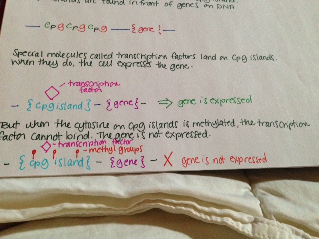
MTHFR mutations and methylation are talked about a lot in the mast cell community. Many people believe that having an MTHFR mutation severely impacts folate metabolism, which in turn means there is not enough methylation, and this dysregulation causes overexpression of genes causing disease. I have searched thoroughly for a link. Really thoroughly. I cannot find any link that is not the idea of one person and researched by that one person, usually outside of peer reviewed settings. I cannot find any link that is not described in detail by a person who does not stand to gain financially from patients who share their beliefs. I am not saying that MTHFR is definitely not linked to mast cell disease. I’m saying I can’t find any proof that it is. I can’t even find anything that SUGGESTS that it is. Might people with these mutations feel better with appropriate folic acid supplementation? Probably. Is that the same thing as causing mast cell disease? Certainly not. It is certainly not the same thing.

I think personal stories hold a lot of power. If your personal story is that your mast cell disease is significantly better controlled by addressing your MTHFR mutation, then I think that is fantastic. I think it is entirely possible that this is the case for many. But I do not believe it causes mast cell disease. And I think everyone would feel better with appropriate levels of folate, as it is an important player in many vital reactions.


References:

Schneider JA, Rees DC, Liu YT, Clegg JB (May 1998). “Worldwide distribution of a common methylenetetrahydrofolate reductase mutation”. Am. J. Hum. Genet. 62 (5): 1258–60

M L Slattery, et al. Methylenetetrahydrofolate reductase, diet and risk of colon cancer. Cancer Epidemiol Biomarkers Prev., 8 (1999), PP 560S-564S.

House, AA; Eliasziw, M; Cattran, DC; Churchill, DN; Oliver, MJ; Fine, A; Dresser, GK; Spence, JD (Apr 28, 2010). “Effect of B-vitamin therapy on progression of diabetic nephropathy: a randomized controlled trial.”. JAMA: the Journal of the American Medical Association 303 (16): 1603–9.

Lonn, E; Yusuf, S; Arnold, MJ; Sheridan, P; Pogue, J; Micks, M; McQueen, MJ; Probstfield, J; Fodor, G; Held, C; Genest J, Jr; Heart Outcomes Prevention Evaluation (HOPE) 2, Investigators (Apr 13, 2006). “Homocysteine lowering with folic acid and B vitamins in vascular disease.”. The New England Journal of Medicine 354 (15): 1567–77

Skibola CF, Smith MT, Kane E, Roman E, Rollinson S, Cartwright RA, Morgan G’ (October 1999). “Polymorphisms in the methylenetetrahydrofolate reductase gene are associated with susceptibility to acute leukemia in adults”. Proc. Natl. Acad. Sci. U.S.A. 96 (22): 12810–5.

Kim, Y I, et al. Methylenetetrahydrofolate reductase polymorphisms, folate and cancer risk: a paradigm of gene-nutrient interactions in carcinogenesis. Nutr Rev 58 (2000), pp 205-209.

Crider, Krista, et al. Folate and DNA Methylation: A Review of Molecular Mechanisms and the Evidence for Folate’s Role. Adv Nutr January 2012 Adv Nutr vol. 3: 21-38, 2012

Xia, Lei-Zhou, et al. Methylenetetrahydrofolate reductase C677T and A1298C polymorphisms and gastric cancer susceptibility. World J Gastroenterol. Aug 28, 2014; 20(32): 11429–11438.

J Chen, et al. MTHFR polymorphism, methyl-replete diets and the risk of colorectal carcinoma and adenoma among US men and women: an example of gene–environment interactions in colorectal tumorigenesis. J. Nutr., 129 (1999), pp. 560S–564S

DNA Methylation: How it works

DNA methylation is one of the ways your cells control which genes to express. It is an example of epigenetic modification. Epigenetics mechanisms like this do not change the DNA sequence, only the way the genes are expressed. Whether or not DNA methylation is heritable is not clear.

This is how DNA methylation works:

  • Cytosine is a nucleotide, a DNA building block.
  • Through the action of an enzyme called methyltransferase, a methyl group is added to cytosine.
  • This is one of the ways your cells know which genes to express.
  • Cytosine is often found next to guanine, another building block. This is sometimes shown in literature as “CG.”
  • Cytosine and guanine are connected by a phosphate group. This is sometimes shown in literature as “CpG.”
  • A bunch of CpG sites together is called a CpG island. These islands are found in front of genes on DNA.
  • Special molecules called transcription factors land on CpG islands. When they do, the cell expresses the gene.
  • But when the cytosine on CpG islands is methylated, the transcription factor cannot bind. The gene is not expressed.

See pictures below.

Methylation is known to have an important role in cancer biology. Methylation of tumor suppressor genes causes the tumor suppressors not to be expressed, resulting in cancer.

Methylation 1Methylation 2Methylation 3

Genetics of MCAS: mutations and methylation

Despite having largely the same symptom profile as SM patients, people with MCAS often lack the signature D816V mutation, considered by many to be a marker of clonality and thus proliferation.  Despite the fact that their mast cells may look normal under a microscope, there is now a growing body of evidence indicating that mast cells in MCAS patients behave aberrantly due to mutations aside from D816V. 
In three studies that look at KIT mutations in MCAS patients, they were found in 26.5%, 44% and 65% of patients, respectively.  Even the average of these three values represents a significant number of people with MCAS who have KIT mutations.  Of note, these mutations are mostly outside of exon 17, where the D816V mutation is found.  In one patient, a mutation was found in the NLRP3 gene, associated with the inflammatory response. 
There are a number of other mutations in genes with various functions commonly found in patients with SM.  These include genes that make proteins to regulate other genes and genes that affect how we make proteins (epigenetic regulatory proteins, splicing machinery and transcription factors.)  To date, there have been no studies looking at whether MCAS patients have similar mutations.  However, there are clear hints that they do. 
The mutations previously mentioned were all somatic and not germline.  This means that the mutations arose after early embryonic development and thus were not heritable.  This fact indicates clearly that there are other germline mutations not yet identified that may induce the subsequent mutations.  This has been bolstered by a 2013 paper that found familial clustering in MCAS patients.   
An important finding is that nearly all mutations found in MCAS patients are heterozygous, meaning only one of the two copies was mutated.  This implies that homozygous mutations, in which both copies are mutated, could cause the cell to die.  Alternatively, the various mutations may work together to make the mutations work as strongly as if there were two mutated copies. 
At CpG sites, the cell can add a special marker to the cytosine called a methyl group.  This is called methylation.  If the cytosine in a gene is methylated, it turns the gene off.  When a gene is turned off, your body will not use that gene or make a protein from it.  This is one type of gene regulation (epigenetics.) 
Looking at the methylation status in the genomic DNA of white blood cells from MCAS patients shows aberrant methylation patterns.  The genes incorrectly methylated included some involved in DNA/RNA repair, DNA/RNA processing, cell death, cell activity and communication with other cells.  195 individual CpG sites have been identified as candidates as markers for MCAS.  Importantly, there is a correlation between the age of symptom onset and the year of birth, which indicates a sort of anticipation of developing MCAS.  This means that gene regulation by methylation could affect acquisition of later, non-heritable mutations like the ones seen in KIT.
A gene is made up of introns and exons.  When an RNA code is made from a gene to tell the cell how to make a protein, the cell cuts out some pieces of that RNA.  These pieces are called introns.  The remaining pieces, which are connected back together, are called exons.  The way the cell cuts the RNA and reconnects the pieces is called splicing.   The mutations in KIT seen in MCAS patients almost all involve intron and exon junctions, where they meet.  There are also some differences in the way splicing occurs in MCAS patients. 

Reference:
Molderings, Gerhard J.  The genetic basis of mast cell activation disease – looking through a glass darkly.  2014.  Critical Reviews in Oncology/Hematology.

Gene expression and the D816V mutation

What exactly is the D816V mutation and why does it matter?  To answer that, we need to understand the basic pathway by which a cell expresses a gene.    

DNA (deoxyribonucleic acid) is the molecule that contains the genetic code for all known living organisms and some viruses.  DNA is composed of two strands that wrap around each other in a double helix pattern.  DNA is built out of nucleotides, molecules that contain energy.  The nucleotides that build DNA are adenine (A), guanine (G), thymine (T) and cytosine (C).  These nucleotides bond in specific pairs.  This means that when one nucleotide in on one strand of DNA, there is a specific nucleotide on the other strand.  A and T, and C and G specifically bond to each other.  They are known as base pairs.  DNA strands made up of base pairs are said to be “complementary.” 


RNA (ribonucleic acid) is a more versatile nucleic acid that codes, regulates and expresses genes, amongst other things.  It also has base pairs: adenine and uracil (U), and thymine and cytosine.  These nucleotides can be complementary to DNA nucleotides.  For example, an RNA adenine is complementary to a DNA thymine, and so on.

DNA replication is the process by which an exact copy of a piece of DNA is made.  This happens when a cell divides.  In replication, the DNA double helix “unzips,” or splits apart into two strands, the base pairs of which are not connected.  Special enzymes move along each of the two split strands and place the appropriate nucleotides next to each strand to form base pairs.  The end result of this is two double helices of DNA that are exact copies.   


Some parts of DNA, called genes, tell the cell how to make proteins or RNA that has a specific function.  (Sometimes RNA can also do this.)  Genes tell the cell how to build and maintain the cell and allow it pass on traits to offspring.  These proteins or RNA are made by expressing the gene.  In gene expression, the information from the gene is turned into a “gene product,” that will be made into something useful for the cell.
Transcription is the start of gene expression.  Gene expression is very complicated and controlled by many mechanisms.  Having a gene does not mean it will always be expressed.  In transcription, a piece of DNA is copied into a complementary RNA strand.   This RNA is called messenger RNA (mRNA.)  This is a complicated process with several steps.  Once a gene is translated, the mRNA with the gene code goes to the ribosome, a place in the cell that makes proteins.  Proteins are made of amino acids. 

So how exactly does the DNA code for the protein the ribosome will make? Let’s focus on that.
The ribosome reads the messenger RNA made from the DNA gene three nucleotides at a time. Again, when using the code to build a protein, the ribosome reads the code in blocks of three nucleotides. These blocks of three nucleotides are called “codons.” Every combination of three-nucleotides tells the ribosome to add a specific amino acid to the protein. The majority of genes are encoded using this same codon code. So by knowing the DNA sequence, we can anticipate the amino acids that build the protein encoded by the gene. 


How does the ribosome know where to start?  There’s a start codon.  (And some other things also.)
How does the ribosome know where to stop?  There’s a stop codon.  (And some other things also.)
There are several types of genetic mutations, or alterations of the code from the one seen in most of the population.  In a point mutation, a single nucleotide is changed.  The D816V mutation is a point mutation. 
We use a specific nomenclature to describe genetic mutations.  Amino acids are often referred to with single letter codes for the sake of brevity.  The amino acid aspartic acid is referred to as “D,” while the amino acid for valine is referred to as “V.”  In the CKIT gene, the amino acid sequence Asp-Phe-Gly (aspartic acid – phenylalanine – glycine) is very important to the receptor being shaped the right way. 
Aspartic acid is encoded by the RNA code “GAU” or “GAC.”  In cells with the D816V mutation, this sequence is changed to “GUU” or “GUC.”  The second base is changed from an A to a U.  Doing this changes the amino acid encoded from aspartic acid (D) to valine (V).  These amino acids are shaped differently, and because of this, the receptor is shaped differently and behaves differently.  When the receptor is made with the amino acid aspartic acid in that place, SCF (stem cell factor) binds to the receptor and activates the cell, telling it not to die and to make more cells.  When the receptor is made with the amino acid valine in that place, the receptor activates itself and SCF is not needed.  It basically tells itself not to die and to make more cells repeatedly. 
So the term “D816V” means that at codon 816, the code was altered in a way that changed the amino acid for aspartic acid to valine.  Some people with mast cell disease don’t have the D816V mutation, but often they have another mutation at codon 816, like D816G.  Sometimes they have a mutation somewhere else in the same “exon.”  An exon is the part of the code that is sent as RNA to be made into a gene product.  The location of the CKIT gene is referred to as exon 17.  

Image credits:


http://www.brooklyn.cuny.edu/bc/ahp/BioInfo/TT/TscriptD.html

http://genmed.yolasite.com

http://www.bristol.k12.ct.us/

http://en.wikipedia.org/wiki/Gene_expression