Skip to content

diagnosis

Lyme Disease: CDC Recommended Diagnostics (part 2)

Diagnosis of Lyme disease is a complicated affair. Part of the complication is the way our immune system responds to Lyme disease, and part of it is the current tests available to test for infection.

An antigen is anything that generates an immune response inside your body. Borrelia spp., the causative agents of Lyme disease, are antigens. They enter your body, your immune system recognizes it as a threat, and generates a specific IgM in response to Borrelia. This IgM is present in your serum about a week after infection, and persists in good quantities until about 12 weeks after infection, with concentration of specific IgM peaking around 4-6 weeks.

About 4 weeks after infection, your body generates Borrelia spp. IgG. This change from making IgM to IgG is called seroconversion. This IgG persists in good quantities for about 9-10 months after infection. While both IgM and IgG titers decrease over time, they can persist in serum for years after infection is resolved.

The standard Lyme diagnostics look for IgM and IgG. To be honest, I was pretty surprised at the abundance of different Lyme diagnostics, including others that look at that antigen and PCR based tests. There are multiple types of tests, and for each type of test, there are multiple companies that make them. It is a bit of a hot mess. So let’s look at the CDC recommended testing first, and then I’ll get to the others.

The CDC recommends a two step process for diagnosing Lyme. The first step is an ELISA and the second is a Western blot.

The Lyme ELISA (enzyme linked immunoassay) tests look for IgM and/or IgG, the antibodies made in response to the infection. These tests are sensitive, meaning that almost everyone with Lyme will test positive IF TESTED FOUR WEEKS AFTER SYMPTOMS PRESENT. (Note: I saw the phrase “almost everyone with Lyme will test positive” in relation to ELISA testing for Lyme several times. This test is most likely to correctly identify positives after patients have seroconverted.) However, these tests are not that specific, so they will provide 5-7% false positives. So with this test, if you have Lyme, you are likely to get a positive result, but if you don’t have it, you might still get a positive result. If the ELISA test is positive, then blot testing is done to confirm the result. If the ELISA test is negative, then the blot is not run, and the patient is told they do not have Lyme disease.

Western blots look for proteins specific to the organism in question. Western blots for Lyme testing are usually called immunoblots. These tests look for the specific IgG and IgM. Like the ELISA, the blot for IgG is more accurate, but requires the patient to have seroconverted. Blots for IgM are more likely to give false positives, so people who don’t have Lyme disease may have a positive test. The CDC guidelines recommend only running IgM blots in the first four weeks of illness due to the higher risk of false positive.

A Western blot works by showing “bands” to demonstrate that particular antigens were found. The bands are visible to the person reading the result. In order for an IgM blot to be considered positive, two of three possible bands must be positive. In order for an IgG blot to be considered positive, five of ten possible bands must be positive.


Let’s review.

You have had symptoms for two weeks. Your IgM ELISA is positive. Your IgG ELISA is negative (because your body hasn’t made IgG yet for Borrelia spp.) Your IgM blot shows 3/3 bands. Your IgG blot is negative. You are diagnosed with Lyme disease.

You have had symptoms for two weeks. Your IgM ELISA is positive. Your IgG ELISA is negative (because your body hasn’t made IgG yet for Borrelia spp.) Your IgM blot shows 1/3 bands. Your IgG blot is negative. You are not diagnosed with Lyme disease.

You have had symptoms for two weeks. Your IgM ELISA is negative. Your IgG ELISA is negative (because your body hasn’t made IgG yet for Borrelia spp.) No blots are run. You are not diagnosed with Lyme disease.

You have had symptoms for six weeks. Your IgM ELISA is positive. Your IgG ELISA is positive. Your IgM blot shows 2/3 bands. Your IgG blot shows 5/10 bands. You are diagnosed with Lyme disease.

You have had symptoms for six weeks. Your IgM ELISA is negative. Your IgG ELISA is positive. Your IgM blot is negative. Your IgG blot shows 5/10 bands. You are diagnosed with Lyme disease.

You have had symptoms for six weeks. Your IgM ELISA is negative. Your IgG ELISA is positive. Your IgM blot is positive. Your IgG blot shows 4/10 bands. You are not diagnosed with Lyme disease.

You have had symptoms for six weeks. Your IgM ELISA is positive. Your IgG ELISA is negative. Your IgM blot shows 3/3 bands. Your IgG blot shows 4/10 bands. You are not diagnosed with Lyme disease.


So let’s talk about this two step process. It has been studied, a lot. The two step process was implemented in the mid-90’s due to the lack of specificity, meaning that people were being treated for Lyme disease who didn’t have it. When the two step process is run and interpreted by skilled operators, according to recommendations, it has 99% specificity. This means a 1% chance of calling someone negative when they really have Lyme disease.

But this doesn’t seem to translate well to clinical practice. A key weakness of these diagnostics in practice is their inability to correctly diagnose people who are newly infected. One study found that the sensitivity and specificity for the two tier test was 100% and 99% for people who no longer had the bull’s eye rash, which happens during the acute phase. But it only had 29% sensitivity for patients in the acute phase with the rash. (Steere 2008) Another study looking particularly at clinical practice found that 50/182 patients had a false positive IgM blot. 78% of them received unnecessary antibiotics. (Seriburi 2012)

I understand why these tests work fine in some studies and not in others. As far as ELISAs giving false positives, that’s no surprise to anyone who has run them before. They’re “sticky.” They crossreact with a lot of things. The reagents are optimized to minimize this, but it is a persistent problem with this type of test. Instead of getting an exact match for your target (Borrelia spp.), you get a “close enough.” You get a positive result if you get a close enough match, and things that match close enough include antibodies for anaplasmosis, leptospirosis, syphilis, Epstein Barr virus, and lupus, among others.

Western blots are also problematic. They are time consuming and complicated. There are a lot of steps and there is a lot of repetition. Reading the results can be confusing. You are comparing the intensity of a band to a control. So let’s say the darker it is, the more positive it is. Just seeing a band does not make it positive. It has to also be as dark as the control for the same band. And things like darkness are subjective.

A sticking point for a lot of people seems to be the fact that they have some bands on their Western blots but not enough to meet the positive criteria. A lot of people feel that if they are positive for some bands that this should be sufficient for a diagnosis. I understand why this is confusing. It’s confusing because it seems like if these blots test for proteins specific to Lyme disease that having any of them should be indicative of infection. But that’s not true.

Remember how I said ELISA tests are sticky, that you can get a good enough match that’s not specific? There’s some of that happening in Western blotting, too. And remember how I said that the procedure is difficult, that the results are subjective? Those facts mean that you can get a few bands through operator error. That’s why it is so important that ALL the required bands show up.

Diagnostic development involves employing a lot of measures to ensure you compensate for errors borne out of biologic crossreaction (like sticky antibodies) and procedural inadequacies. The result is very specific criteria for what is considered positive and what is not. The FDA requires this.

Another thing to keep in mind for later is that diagnostics that are FDA validated are designed for very particular uses. That means that if you have an FDA validated test for Lyme IgG in serum and you use that test to test whole blood or cerebrospinal fluid or synovial fluid, and you get a positive result, it is meaningless. It is not positive. It is nothing. If you have an FDA validated test that tells you to use 100 ul of serum and 100 ul of something else per tube, and you use 150 ul of serum and 150 ul of something else per tube, and you get a positive result, it is meaningless. This also generates a lot of confusion because people feel like if you are looking for the same thing (Lyme IgG), it shouldn’t matter where you find it. But it does.

It matters because sample matrices (like serum or whole blood or whatever) have their own specific “backgrounds.” This means that they contain different proteins and have different fluid dynamics and they could cause additional crossreaction or they could cause less crossreaction. The chemistry involved in diagnostics is very fine. Tests that have a large tolerance for changes like this are called “robust.” The Lyme diagnostics discussed here are not robust.


References:

Steere AC, McHugh G, Damle N, Sikand VK. Prospective study of serologic tests for Lyme disease. Clin Infect Dis. 2008;47(2):188–195. doi: 10.1086/589242.

Wormser GP, Carbonaro C, Miller S, Nowakowski J, Nadelman RB, Sivak S, Aguero-Rosenfeld ME. A limitation of 2-stage serological testing for Lyme disease: enzyme immunoassay and immunoblot assay are not independent tests. Clin Infect Dis. 2000;30(3):545–548. doi: 10.1086/313688

Klempner, M. S., C. H. Schmid, L. Hu, A. C. Steere, G. Johnson, B. McCloud, R. Noring, and A. Weinstein. 2001. Intralaboratory reliability of serologic and urine testing for Lyme disease. Am. J. Med. 110:217-219

Reed, Kurt D. Laboratory Testing for Lyme Disease: Possibilities and Practicalities. J Clin Microbiol. Feb 2002; 40(2): 319–324.

Ang, C. W., et al. Large differences between test strategies for the detection of anti-Borrelia antibodies are revealed by comparing eight ELISAs and five immunoblots. Eur J Clin Microbiol Infect Dis. Aug 2011; 30(8): 1027–1032.

Woods, Charles R. False-Positive Results for Immunoglobulin M Serologic Results: Explanations and Examples. J Ped Infect Dis (2013).

Seriburi, N. Ndukwe, Z. Chang, M. E. Cox and G. P. Wormser . High frequency of false positive IgM immunoblots for Borrelia burgdorferi in Clinical Practice. Clin Microbiol Infect 2012; 18: 1236–1240

Branda, John A, et al. 2-Tiered Antibody Testing for Early and Late Lyme Disease Using Only an Immunoglobulin G Blot with the Addition of a VlsE Band as the Second-Tier Test. Clin Infect Dis. (2010) 50 (1): 20-26.
Progression of mast cell diseases (Part 3)

What causes aberrant mediator release in mast cell activation diseases (including MCAS and SM)?

“Selective release of mediators during mast cell activation may be accomplished in three important and possibly interrelated ways. One is by activation via one of the mast cell’s non-IgE receptors, for instance, through the activation of the IL-1 receptor… Another way in which mast cells may selectively activate is through ‘piecemeal’ release of mediators stored in the secretory granules (such as histamine and serotonin)… Lastly, downstream signaling pathways may affect mast cell activation… Differential activation of mast cells in any of these ways may clinically manifest as nc-MCAS.” (Cardet 2013)

“It is also conceivable that mast cells in this group of patients may aberrantly possess a lower threshold to release mediators… It is also conceivable that patients with nc-MCAS are symptomatic because of an abnormal tissue response to physiologically appropriate release of MC mediators.” (Cardet 2013)

“The mutations underlying systemic MCAD drive aberrant mediator production/release with or without readily histologically detectable mast cell accumulation. Mast cell accumulation is due predominantly to a decrease in mast cell apoptosis (refs 30,31 and further references therein). On a limited scale, it is also due to an increase in proliferation.” (Haenisch 2012)


Do all SM patients have elevated n-methylhistamine and prostaglandin F2a?

71% had elevated urinary histamine in 24 hr test; 81% had elevated urinary n-methylhistamine in 24 hr test; 75% had elevated urinary PGF2a in 24 hr test. (Lim 2009)


If my tryptase is normal, does that mean I don’t have SM?

In patients tested, 96% had elevated tryptase over 11.5 ng/ml. (Lim 2009)

“20% to 30% of SM patients have serum tryptase levels below the WHO-defined threshold of 20 ng/mL (sensitivity 80%, specificity 98%).” (Pardanini 2013)


If my blood test for the D816V mutation is negative, I definitely don’t have it, right?

“The sensitivity of KITD816V detection in peripheral blood is suboptimal, and tests for non-KITD816V mutation screening may not be readily available.” (Pardanini 2013)

“I prefer using DNA from BM aspirate for KITD816V screening given the low sensitivity of peripheral blood in this regard… Using this approach, we found 78% of ISM patients to harbor KITD816V.” (Pardanini 2013)

“Although, the sensitivity of KITD816V detection may be higher when using sorted or purified mast cells, this option is not routinely available. Consequently, the inability to detect KITD816V in peripheral blood does not exclude SM [].” (Pardanini 2013)


How often do SM patients not meet the major diagnostic criteria (mast cell aggregates)?

“Attempts at validating the WHO diagnostic criteria reveal that approximately 20% of ISM patients lack mast cell clusters in the BM and approximately 30% exhibit a serum tryptase level < 20 ng/mL.” (Sanchez 2011)


Is MCAS the same as HIT (histamine intolerance)?

“[S]ome have proposed that a deficiency in the enzymes responsible for histamine metabolism, diamine oxidase (DAO) and histamine N-methyltransferase, leads to excess levels of histamine and therefore histamine intolerance, with clinical manifestations not unlike those described for nc-MCAS… There is no scientific literature to support their relevance to nc-MCAS.” (Cardet 2013)


Are MCAS patients usually positive for the three most commonly tested mediators (tryptase, n-methylhistamine, PGD2?)

“Although all of our patients with MCAS had a positive test result for at least 1 MC mediator, only 33%, 56%, and 44% of the patients had positive test results for tryptase, histamine, and PGD2, respectively.” (Hamilton 2011)


Will my MCAS symptoms ever get better?

“Most patients with MCAS in our cohort who were treated with anti-MC mediator medications responded dramatically. After an average of 4.6 years of MC-related symptoms, 66% of the patients with MCAS achieved a complete or major regression in symptoms to MCAS treatment.” (Hamilton 2011)

“It is important to mention that no defining characteristics (eg, presence of allergies or history of anaphylaxis) could be identified that distinguished those who had a complete regression in symptoms versus those who did not.” (Hamilton 2011)

“The most impressive treatment responses were for abdominal pain (14/17 of the patients who initially had the symptom responded), headache (12/15), poor concentration and memory (7/12), and diarrhea (9/12); there was a more modest response to flushing (6/16). We also found that all but 1 of our patients with MCAS had a sustained response to anti-MC mediator medications. Patients in our cohort were followed for an average of 2.8 years (range, 1-4 years).” (Hamilton 2011)

“In patients with MCAS the rate of response to antimediator therapy is rather good, with 33% showing complete response, 33% a major response, and 33% a minor response after 1 year of treatment.” (Picard 2013)


How prevalent is MCAS?

“MCAS seems to be a more common disorder. Evidence has been presented that MCAS may be an underlying cause of various clinical presentations, e.g. in subsets of patients with fibromyalgia and irritable bowel syndrome. Hence, the prevalence of MCAS is likely to lie within the single-digit percentage range.” (Haenisch 2012)

“Mast cell activation disease in general has long been thought to be rare. However, although SM and MCL as defined by the WHO criteria are truly rare, recent findings suggest MCAS is a fairly common disorder. Evidence has been presented for a causal involvement of pathologically active mast cells not only in the pathogenesis of SM and MCAS but also in the etiology of idiopathic anaphylaxis, interstitial cystitis, some subsets of fibromyalgia and some subsets of irritable bowel syndrome.” (Molderings 2011)


References:

Juan-Carlos Cardet, Maria C. Castells, and Matthew J. Hamilton. Immunology and Clinical Manifestations of Non-Clonal Mast Cell Activation Syndrome. Curr Allergy Asthma Rep. Feb 2013; 13(1): 10–18.

LimKH, TefferiA, LashoTL, et al. Systemic mastocytosis in 342 consecutive adults: survival studies and prognostic factors. Blood 2009; 113(23): 5727-5736.

Britta Haenisch, Markus M. Nothen and Gerhard J. Molderings. Systemic mast cell activation disease: the role of molecular genetic alterations in pathogenesis, heritability and diagnostics. Immunology 2012, 137, 197–205.

Animesh Pardanani. How I treat patients with indolent and smoldering mastocytosis (rare conditions but difficult to manage.) April 18, 2013; Blood: 121 (16).

Matthieu Picard, Pedro Giavina-Bianchi, Veronica Mezzano, Mariana Castells. Expanding Spectrum of Mast Cell Activation Disorders: Monoclonal and Idiopathic Mast Cell Activation Syndromes. Clinical Therapeutics, Volume 35, Issue 5, May 2013, Pages 548–562.

Gerhard J Molderings, Stefan Brettner, Jürgen Homann, Lawrence B Afrin. Mast cell activation disease: a concise practical guide for diagnostic workup and therapeutic options. Journal of Hematology & Oncology 2011, 4:10.


I think I might have mast cell disease: FAQ

What kinds of symptoms do mast cell patients have?

Mast cell disease can cause a variety of symptoms. Each person has their own unique constellation of complaints, and they can vary from day to day. Mast cell patients often have allergic type reactions to many things. They may have had anaphylaxis in the past, but that is not always the case.

What kind of doctor should I see if I think I have mast cell disease?

Due to the fact that mast cell disease can affect multiple body systems, it is managed by doctors of multiple disciplines. Immunologists, dermatologists, gastroenterologists and hematologists/ oncologists all may treat mast cell disease. It really depends who is familiar with mast cell disease in your areas. Immunologists are often the first stop for patients investigating mast cell disease.

Will any doctor know about mast cell disease?

No. Mast cell disease is uncommon. Many doctors are only aware of the types associated with pathologic rashes (cutaneous mastocytosis) or proliferation of mast cells in the bone marrow (systemic mastocytosis.)

How do I get determine if I have mast cell disease?

Labs for diagnosing mast cell disease include serum tryptase (a blood test), n-methylhistamine (24 hour urine test) and D2/F2a prostaglandin (24 hour urine test.) These tests are time sensitive for many patients and have special handling in most labs. Depending on these results, a bone marrow biopsy may be needed.

Can I have mast cell disease if my tryptase is normal?

Yes. 15% of patients with systemic mastocytosis have normal tryptase levels, and the majority of MCAS patients have normal tryptase levels.

How is mast cell disease treated?

Treatment generally focuses on the symptoms. The most common treatments include antihistamines, leukotriene inhibitors and mast cell stabilizers.

Will I feel better with treatment?

Most people feel better with treatment than without, but how much each person recovers is individual. Lifestyle modifications and medications can help many people live a full life.

Is mast cell disease curable?

No. Patients may have a remission from symptoms, but they will always have mast cell disease.

MCAS: Differing criteria among experts

What criteria you have to meet to be diagnosed with MCAS depends on which doctor you see – even the experts don’t agree.

Molderings, Afrin 2011 Akin, Valent, Metcalfe 2010 Valent, Akin, Castells, Escribano, Metcalfe et al 2012
MCAD (umbrella term including both MCAS and SM) diagnosed if both major criteria, or one major criterion and one minor criterion, are present; following bone marrow biopsy, diagnosis is narrowed down to either SM or MCAS MCAS diagnosed if all criteria are met MCAS diagnosed if all criteria are met

Major Criteria

Multifocal of disseminated dense infiltrates of mast cells in bone marrow biopsies and/or in sections of other extracutaneous organ(s) (GI tract biopsies; CD117-, tryptase- and CD25- stained)
Episodic symptoms consistent with mast cell mediator release affecting ≥2 organ systems evidenced as follows:
  1. Skin: urticaria, angioedema, flushing
  2. Gastrointestinal: nausea, vomiting, diarrhea, abdominal cramping
  3. Cardiovascular: hypotensive syncope or near syncope, tachycardia
  4. Respiratory: wheezing
  5. Naso-ocular: conjunctival injection, pruritus, nasal stuffiness
Typical clinical symptoms
Unique constellation of clinical complaints as a result of a pathologically increased mast cell activity (mast cell mediator release symptom) A decrease in the frequency or severity or resolution of symptoms with antimediator therapy: H1– and H2-histamine receptor inverse agonists, antileukotriene medications (cysteinyl leukotriene receptor blockers or 5-lipoxygenase inhibitor), or mast cell stabilizers (cromolyn sodium) Increase in serum total tryptase by at least 20% above baseline plus 2 ng/ml during or within 4 h after a symptomatic period
  Evidence of an increase in a validated urinary or serum marker of mast cell activation: documentation of an increase of the marker to greater than the patient’s baseline value during a symptomatic period on ≥2 occasions or, if baseline tryptase levels are persistently >15 ng, documentation of an increase of the tryptase level above baseline value on 1 occasion. Total serum tryptase level is recommended as the marker of choice; less specific (also from basophils) are 24-hour urine histamine metabolites or PGD2 or its metabolite 11-β-prostaglandin F2. Response of clinical symptoms to histamine receptor blockers or MC-targeting agents e.g. cromolyn
  Rule out primary and secondary causes of mast cell activation and well-defined clinical idiopathic entities

Minor Criteria

Mast cells in bone marrow or other extracutaneous organ(s) show an abnormal morphology (>25%) in bone marrow smears or in histologies
Mast cells in bone marrow express CD2 and/or CD25
Detection of genetic changes in mast cells from blood, bone marrow or extracutaneous organs for which an impact on the state of activity of affected mast cells in terms of an increased activity has been proved
Evidence of a pathologically increased release of mast cell mediators by determination of the content of:

  1. Tryptase in blood
  2. N-methylhistamine in urine
  3. Heparin in blood
  4. Chromogranin A in blood
  5. Other mast cell specific mediators (leukotrienes, PGD2)

Mast cell mediators: Recommended testing for MCAS diagnosis

Lab tests specific to mast cell activation for suspected MCAS patients should include serum tryptase, serum chromogranin A, plasma histamine, chilled plasma PGD2, stat chilled plasma heparin, chilled urine for PGD2, PGF2a and n-methylhistamine. 
Tryptase is the most famous mast cell mediator.  It is a complex molecule with many functions in the body.  It is easily damaged by heat and has a short half-life in the body (6-8 minutes in health subjects, 1.5-2.3 hours in patients with hypersensitivity reactions.  In separated serum, it can last approximately four days.  Serum tryptase value is usually normal in MCAS patients, but sometimes it is elevated.  Tryptase values that show an increase of 20% + 2 ng/ml above the baseline level are considered diagnostic for MCAS.
Chromogranin A is a heat-stable mast cell mediator.  High levels can suggest MCAS, but other sources must first be ruled out, such as heart failure, renal insufficiency, neuroendocrine tumors and proton pump inhibitor (PPI) use.  Starting or stopping PPI therapy will generally cause a change in value within five days.  Once other causes have been excluded, serum chromogranin A can be considered a reliable marker of mast cell activity. 
Heparin is a very sensitive and specific marker of mast cell activation.  However, due to its quick metabolism in the body, it is very difficult to measure reliably.  It has a very short half life and quickly deteriorates, even when refrigerated.  Values above 0.02 anti-Factor Xa units/ml are abnormal, but many commercial tests cannot test that low.  Elevated plasma heparin is sometimes found in MCAS patients. 
Histamine is also released by basophils, but the majority is released by mast cells.  It is heat stable and has a short half life in the body.  Serum histamine peaks at about 5 minutes after release and returns to baseline within 15-30 minutes in most patients.  In separated plasma, it is stable at room temperature for at least 48 hours.  It is broken down to n-methylhistamine, which is more stable and can be measured accurately longer.  N-methylhistamine is usually measured in a 24 hour urine test to account for the variability in release over the course of the day. 
Prostaglandin D2 is produced by several other cell types, but mast cell release is responsible for the dominant amount found in the body.  MCAS patients typically produce much higher levels of PGD2 than n-methylhistamine.  PGD2 is less stable than histamine, being metabolized completely in an estimated 30 minutes.  Its metabolite, PGF2a, is the preferred compound for detection due to its superior stability.    Accurate prostaglandin testing relies upon refrigeration of the sample from the start of collection through testing.  NSAIDs inhibit prostaglandin production and can lower PGD2 in blood and urine.  Renal insufficiency may produce an inaccurately low test value, but elevated prostaglandins are sometime seen in patients with renal disease.  Prostaglandins D2 and F2a can be tested in serum, but 24 hour urine samples are considered more accurate.

Leukotriene B4 and cysteinyl leukotrienes C4, D4 and E4 have been noted to be elevated in SM patients and during acute asthma attacks.  Though commercial testing for these compounds is not easily accessible, but they may be elevated in MCAS patients as well.  Other less specific mast cell mediators that are sometimes abnormal in MCAS patients include Factor VIII, plasma free norepinephrine, tumor necrosis factor alpha, and interleukin-6.

References:
Sur R, Cavender D, Malaviya R. Different approaches to study mast cell functions.  Int. Immunopharmacol. 2007 May;7(5):555-567.
Pregun I, Herszényi L, Juhász M, Miheller P, Hritz I, Patócs A, Rácz K, Tulassay Z. Effect of proton-pump inhibitor therapy on serum chromogranin A level. Digestion 2011; 84:22-28.
Seidel H, Molderings GJ, Oldenburg J, Meis K, Kolck UW, Homann J, Hertfelder HJ. Bleeding diathesis in patients with mast cell activation disease. Thromb. Haemost. 2011 Nov; 106(5):987-989.
Laroche D, Vergnaud MC, Sillard B, Soufarapis H, Bricard H. Biochemical markers of anaphylactoid reactions to drugs: comparison of plasma histamine and tryptase. Anesthesiol. 1991 Dec; 75(6):945-949.

Takeda J, Ueda E, Takahashi J, Fukushima K. Plasma N-methylhistamine concentration as an indicator of histamine release by intravenous d-tubocurarine in humans: preliminary study in five patients by radioimmunoassay kits. Anesth. Analg. 1995; 80:1015-1017.

Maclouf J, Corvazier E, Wang ZY. Development of a radioimmunoassay for prostaglandin D2 using an antiserum against 11-methoxime prostaglandin D2. Prostaglandins 1986 Jan; 31(1):123-132.
Freeman JG, Ryan JJ, Shelburne CP, Bailey DP, Bouton LA, Narasimhachari N, Domen J, Siméon N, Couderc F, Stewart JK. Catecholamines in murine bone marrowderived mast cells. J. Neuroimmunol. 2001 Oct;119(2):231-238.
Gordon JR, Galli SJ. Mast cells as a source of both preformed and immunologically inducible TNF-α/cachectin. Nature 1990 Jul 19; 346:274-276.

MCAD, MCAS and the hierarchy of mast cell disease classifications

I have seen several posts recently expressing confusion about various mast cell diagnoses so I figured I would put up a post to clear things up.
Mast cell activation disorder (MCAD) is a catch-all term for mast cell disease (MCD.)  MCAD and MCD can be used interchangeably.  So if you have any mast cell disease, you have MCAD.  If you have SM, you have MCAD, because SM is a type of MCAD.  If you have UP, you have MCAD, and so on.  MCAD is an umbrella term.  It is non-specific.  It is similar to being told that you have heart disease when you have mitral valve prolapse.  It is true, but it is not precise enough to give all information needed to treat effectively.
Mast cell activation syndrome (MCAS) is the diagnosis you get if you do not meet the criteria for any of the defined mast cell diseases, but have mast cell mediator related symptoms.  You cannot have MCAS and another mast cell disease because, by its definition, MCAS is ONLY diagnosed if you do NOT meet the criteria for any other mast cell disease.  You cannot have UP and MCAS.  You cannot have SM and MCAS.  I think some people think that MCAS means you have mediator related symptoms.  This is not the case.  You can have mediator related symptoms with pretty much any mast cell disease. 
A paper was published a few years ago by a doctor who considers mast cell activation symptoms to be due exclusively to proliferation (like in SM.)  He wrote a paper that says that MCAS is found in people with SM.  This paper sort of confused the issue for a lot of people.  However, the mast cell community (including researchers and prominent doctors) do not consider this to be the case.  They agree that you cannot have SM and MCAS.
Also confusing is the fact that mast cell activation (MCA) is NOT the same as MCAS.  MCA just means that your mast cells are activated, which occurs in any mast cell disease.  MCA is not a diagnosis, it is a symptom.  So you can have MCA in SM.  But you still can’t have MCAS in SM.
So if you have SM and have lots of mediator related symptoms, you have SM.  If you want to speak broadly, you have SM.
If you test negative for SM and have no CM, but have mast cell symptoms and elevated mast cell markers, you have MCAS. 
If you have UP and then later develop SM, you have SM with skin involvement, or SM with UP. 
If you have UP or TMEP and have lots of mediator related systemic symptoms, you do NOT have UP and MCAS.  You have UP.  UP and TMEP (forms of CM) can cause systemic symptoms.  But you cannot have MCAS because you can only have MCAS if you do not meet the criteria for another mast cell disease.
Let’s review.
If you have UP: you have UP, you have CM, you have MCAD.
If you have TMEP: you have TMEP, you have CM, you have MCAD.
If you have SM: you have SM, you have MCAD.
If you have SM with UP: you have SM with skin involvement, you have UP, you have MCAD.
If you have SM with TMEP: you have SM with skin involvement, you have TMEP, you have MCAD.
If you have SM-AHNMD: you have SM-AHNMD, you have MCAD.
If you have ASM: you have ASM, you have MCAD.
If you have MCL: you have MCL, you have MCAD.

If you have MCAS: you have MCAD.

Reference:
Molderings GJ, Brettner S, Homann J, Afrin LB. Mast cell activation disease: a concise practical guide for diagnostic workup and therapeutic options. J. Hematol. Oncol.2011; 4:10-17.

Diagnosis of mast cell diseases

There seems to be a lot of confusion regarding diagnosis of mast cell diseases, so I figured I’d do a review.

Cutaneous mastocytosis (CM) is diagnosed by skin biopsy.  Urticaria pigmentosa (UP), also called maculopapullar cutaneous mastocytosis (MPCM), diffuse cutaneous mastocytosis (DCM) and telangiectasia macularis eruptive perstans (TMEP) are types of cutaneous mastocytosis.  They each present with a rash and may have accompanying systemic symptoms. 
Mastocytoma of the skin is also diagnosed by skin biopsy.
Systemic mastocytosis (SM) has the following diagnostic criteria:
Major:
1.       Multifocal, dense infiltrates of mast cells (15 or more in an aggregate) detected in sections of bone marrow and/or extracutaneous organ. 
Minor:
1.       In biopsy sections, more than 25% of mast cells in infiltrated area are spindle-shaped or have atypical morphology; or, of all mast cells in bone marrow aspirate smears, more than 25% are immature of atypical. 
2.       Detection of Kit mutation at codon 816 in bone marrow, blood or other extracutaneous organ.
3.       Mast cells in bone marrow, blood or other extracutaneous organ that co-express CD117 with CD2 and/or CD25.
4.       Serum total tryptase persistently >20 ng/mL (if there is not a clonal myeloid disorder.)
SM is diagnosed if a patient has either one major and one minor criteria, or three minor criteria.  So let’s look at how this plays out.
A patient with mast cell symptoms gets a bone marrow biopsy.  It shows more than 25% abnormal mast cells in the section.  They are CKIT negative, have a serum tryptase of 2, and do not express CD2/CD25.  They are diagnosed with SM.
A patient has a biopsy that does not show dense infiltrates.  All their mast cells are shaped normally.  In blood tests, their mast cells are found to express CD2.  They are CKIT+, also from blood.  Their serum tryptase is 28.  They are diagnosed with SM.
A patient has a biopsy that shows dense infiltrates, but they have less than 25% abnormal mast cells and their mast cells do not express CD2/CD25.  They are CKIT- and have a serum tryptase of 18.  They are not diagnosed with SM.
A few things to keep in mind:
Most people with SM are diagnosed by bone marrow biopsy, but a biopsy from any non-skin organ showing mast cell infiltration as described above can be used.  This means if you have a positive lung biopsy, liver biopsy, whatever, you may not necessarily need a bone marrow biopsy. 
It can take up to six bone marrow biopsies to diagnose SM in a patient who has had it the entire time.  This is because there is no way to know where the mast cells will cluster.  A negative bone marrow biopsy does not necessarily mean that you do not have SM.  Hence the minor criteria.
The CKIT test looks for a specific mutation, the D816V mutation.  There are other mutations found in codon 816.  You may have a mutation but test CKIT- because you do not have the D816V mutation.  Also, the blood test for CKIT is not always reliable.  The test way to test this is from a bone marrow sample.  You could test CKIT- in blood and then test CKIT+ in bone marrow.
The serum tryptase criterion refers to persistent baseline level tryptase, not reaction level tryptase. 
So let’s say you have a negative bone marrow biopsy and a blood test that shows you are CKIT+ and have mast cells expressing CD2/CD25.  What do you have?  You have monoclonal mast cell activation syndrome (MMAS.)  MMAS is diagnosed in patients who have one or two of the minor criteria for systemic mastocytosis.
Let’s say you have a negative bone marrow biopsy and blood work that shows normal mast cells and tryptase below 20, but you have systemic symptoms.  What do you have?  You probably have MCAS (mast cell activation syndrome.)  There are some other tests for that.  24 hour urine tests are usually done to measure the levels of histamine metabolites and prostaglandin D2 metabolites.
The following are the diagnostic criteria for MCAS:
1.       Episodic symptoms consistent with mast cell mediator release affecting two or more organ systems: skin (urticarial, angioedema, flushing); GI (nausea, vomiting, diarrhea, cramping); cardiovascular (fainting or near fainting due to low blood pressure, rapid heartbeat); respiratory (wheezing); naso-ocular (itching, nasal stuffiness, red eyes.)
2.       A decrease in frequency or severity; or resolution of symptoms with antihistamines, leukotriene inhibitors or mast cell stabilizers.
3.       Evidence of elevation of urinary or serum marker of mast cell activation: Documentation of elevation of marker during a symptomatic period on at least two occasions, or if baseline tryptase is persistently above 15 ng.  This includes urinary histamine and prostaglandin D2.
4.       Clonal and secondary disorders of mast cell activation ruled out.
MCAS is a diagnosis of exclusion.  It is the diagnosis you receive if you have mast cell symptoms that are ameliorated with mast cell medications if you do not meet the criteria for any other mast cell disease.
Back to SM.  Let’s say you’re positive for SM.  Now what?
They will determine if you have other important markers of disease severity.  These are called B and C findings.  They are as follows:
B findings:
1.       Increased mast cell burden (>30% mast cell aggregates on bone marrow biopsy and/or serum tryptase >200 ng/ml).
2.       Hypercellular marrow, signs of overproduction or abnormal development of blood cells, normal or slightly abnormal blood counts that are not abnormal enough to be considered an associated hematologic disorder.
3.       Swelling of the liver that can be felt manually, no free fluid or signs of dysfunction, persistently swollen glands, swelling of the spleen that can be felt manually without signs of dysfunction.
If you have two or more B findings, you have SSM (smoldering systemic mastocytosis.) 
C findings:
1.       Unusual blood counts (low ANC, low Hb, low platelets)
2.       Swelling of the liver that can be felt manually, with impaired liver function, free fluid and/or portal hypertension.
3.       Large osteolytic lesions and/or pathological fractures.
4.       Swelling of the spleen with impaired function.
5.       Malabsorption with weight loss and/or low albumin.
If you have one or more C finding, you have ASM (aggressive systemic mastocytosis.)
How are these B and C findings identified?  Bone marrow biopsy, blood tests and imaging (ultrasounds, MRI, etc.) 
If you have SM and one B finding, or no B findings, you have indolent systemic mastocytosis (ISM.) 
If your bone marrow biopsy shows significant overproduction or abnormal development of a cell type that is not a mast cell, you may be diagnosed with SM-AHNMD (systemic mastocytosis with associated hematologic non-mast cell lineage disease.)  People with this type of SM also have another blood disorder, such as chronic myelogenous leukemia, myelodysplasia, etc.  In these patients, serum tryptase is not reliable to assess mast cell burden.  
Mast cell leukemia (MCL) is extremely rare.  It is diagnosed by >20% mast cells on the bone marrow aspirate smear.   
Mast cell sarcoma is a very aggressive form of sarcoma.  It is diagnosed by biopsy of the tumor.  People with these tumors quickly developed mast cell leukemia.  There have only been three cases reported in literature.  To be clear, this is NOT the same as mastocytoma.  Mastocytomas are benign.
I think I got everything.  Any questions?  Ask in the comments.

Gene expression and the D816V mutation

What exactly is the D816V mutation and why does it matter?  To answer that, we need to understand the basic pathway by which a cell expresses a gene.    

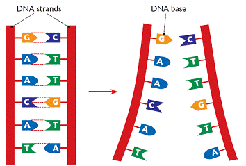
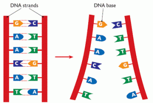
DNA (deoxyribonucleic acid) is the molecule that contains the genetic code for all known living organisms and some viruses.  DNA is composed of two strands that wrap around each other in a double helix pattern.  DNA is built out of nucleotides, molecules that contain energy.  The nucleotides that build DNA are adenine (A), guanine (G), thymine (T) and cytosine (C).  These nucleotides bond in specific pairs.  This means that when one nucleotide in on one strand of DNA, there is a specific nucleotide on the other strand.  A and T, and C and G specifically bond to each other.  They are known as base pairs.  DNA strands made up of base pairs are said to be “complementary.” 


RNA (ribonucleic acid) is a more versatile nucleic acid that codes, regulates and expresses genes, amongst other things.  It also has base pairs: adenine and uracil (U), and thymine and cytosine.  These nucleotides can be complementary to DNA nucleotides.  For example, an RNA adenine is complementary to a DNA thymine, and so on.

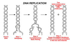
DNA replication is the process by which an exact copy of a piece of DNA is made.  This happens when a cell divides.  In replication, the DNA double helix “unzips,” or splits apart into two strands, the base pairs of which are not connected.  Special enzymes move along each of the two split strands and place the appropriate nucleotides next to each strand to form base pairs.  The end result of this is two double helices of DNA that are exact copies.   


Some parts of DNA, called genes, tell the cell how to make proteins or RNA that has a specific function.  (Sometimes RNA can also do this.)  Genes tell the cell how to build and maintain the cell and allow it pass on traits to offspring.  These proteins or RNA are made by expressing the gene.  In gene expression, the information from the gene is turned into a “gene product,” that will be made into something useful for the cell.
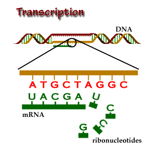
Transcription is the start of gene expression.  Gene expression is very complicated and controlled by many mechanisms.  Having a gene does not mean it will always be expressed.  In transcription, a piece of DNA is copied into a complementary RNA strand.   This RNA is called messenger RNA (mRNA.)  This is a complicated process with several steps.  Once a gene is translated, the mRNA with the gene code goes to the ribosome, a place in the cell that makes proteins.  Proteins are made of amino acids. 

So how exactly does the DNA code for the protein the ribosome will make? Let’s focus on that.
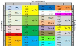
The ribosome reads the messenger RNA made from the DNA gene three nucleotides at a time. Again, when using the code to build a protein, the ribosome reads the code in blocks of three nucleotides. These blocks of three nucleotides are called “codons.” Every combination of three-nucleotides tells the ribosome to add a specific amino acid to the protein. The majority of genes are encoded using this same codon code. So by knowing the DNA sequence, we can anticipate the amino acids that build the protein encoded by the gene. 


How does the ribosome know where to start?  There’s a start codon.  (And some other things also.)
How does the ribosome know where to stop?  There’s a stop codon.  (And some other things also.)
There are several types of genetic mutations, or alterations of the code from the one seen in most of the population.  In a point mutation, a single nucleotide is changed.  The D816V mutation is a point mutation. 
We use a specific nomenclature to describe genetic mutations.  Amino acids are often referred to with single letter codes for the sake of brevity.  The amino acid aspartic acid is referred to as “D,” while the amino acid for valine is referred to as “V.”  In the CKIT gene, the amino acid sequence Asp-Phe-Gly (aspartic acid – phenylalanine – glycine) is very important to the receptor being shaped the right way. 
Aspartic acid is encoded by the RNA code “GAU” or “GAC.”  In cells with the D816V mutation, this sequence is changed to “GUU” or “GUC.”  The second base is changed from an A to a U.  Doing this changes the amino acid encoded from aspartic acid (D) to valine (V).  These amino acids are shaped differently, and because of this, the receptor is shaped differently and behaves differently.  When the receptor is made with the amino acid aspartic acid in that place, SCF (stem cell factor) binds to the receptor and activates the cell, telling it not to die and to make more cells.  When the receptor is made with the amino acid valine in that place, the receptor activates itself and SCF is not needed.  It basically tells itself not to die and to make more cells repeatedly. 
So the term “D816V” means that at codon 816, the code was altered in a way that changed the amino acid for aspartic acid to valine.  Some people with mast cell disease don’t have the D816V mutation, but often they have another mutation at codon 816, like D816G.  Sometimes they have a mutation somewhere else in the same “exon.”  An exon is the part of the code that is sent as RNA to be made into a gene product.  The location of the CKIT gene is referred to as exon 17.  

Image credits:


http://www.brooklyn.cuny.edu/bc/ahp/BioInfo/TT/TscriptD.html

http://genmed.yolasite.com

http://www.bristol.k12.ct.us/

http://en.wikipedia.org/wiki/Gene_expression

Bone marrow biopsy

Most people with suspected systemic mastocytosis receive a bone marrow biopsy as part of their diagnostic testing.  Sometimes people will have confirmed mast cell infiltration in another organ, in which case a bone marrow biopsy may not be needed. 
I know that once I needed a bone marrow biopsy, I sort of felt like my illness had hit the big time.  Like it was time to be really concerned.  My family and friends were really concerned because bone marrow biopsies are used to diagnosis serious diseases.  It is okay to be scared.  But the procedure was not even close to the worst I’ve had, and the pain was manageable. 
In the middle of long bones, there is a squishy center called bone marrow.  Your bone marrow produces most of your body’s blood cells.  The marrow is essentially organized tissue that holds the immature blood cells.
Red bone marrow is active and produces red blood cells, platelets, neutrophils, basophils, eosinophils, monocytes/macrophages, T cells, B cells and mast cells.  Yellow bone marrow mostly contains fat.  Red bone marrow is found in flat bones, like the sternum (breast bone) and the pelvic girdle (upper hip bones.)  In children, the femur (thigh bone) also contains red marrow.
A bone marrow biopsy removes some of the solid tissue from the red marrow to look for abnormal cells.  It uses a long, hollow needle.   Using this needle, a solid piece of bone marrow is removed.  This is called a core. 
A bone marrow aspiration, which removes some liquid from the red marrow, is often done at the same time.  It uses a syringe to remove a little bit of the liquid. 
When you arrive, you may be given IV sedation.  You usually have to request this in advance.  Generally, this is “twilight sedation,” in which you are awake but given medications to relax and manage the pain. 
If they are taking samples from the back of your pelvic crest, you lay on your stomach.  You receive a local anesthetic, typically lidocaine.  It will burn as the drug numbs the area.  (I’ve always found it really ironic that an anesthetic burns.)  A small incision is made in the skin at the biopsy site. 
A hollow needle is then pushed through the bone and into the marrow.  A syringe is attached to the needle and the person doing the procedure pulls back on the plunger to draw liquid into the needle.  This is called the aspirate.  When the aspirate is removed, it changes the pressure inside the bone and causes some pain.  Sometimes no aspirate is found.  This is called a “dry tap.”  If this occurs, another site is biopsied. 
After the aspiration, the biopsy is performed.  This uses a larger hollow needle that is pushed through the bone and into the solid marrow.  The entire procedure (aspiration and biopsy) usually takes about 30 minutes.
After the samples are taken, a sterile dressing is applied to the site with pressure to stop bleeding.  Once the bleeding has stopped, a new dressing is taped into place.  It is important to keep this dressing dry for 24 hours, as getting it wet can increase the risk of infection.  After 24 hours, you can shower or swim as usual. 
The biopsy site will be sore for at least a few days.  Avoid strenuous activity for a few days.  If you develop an (unusual) fever, severe pain, swelling, redness or drainage from the site, or uncontrolled bleeding, contact your health care provider.  This can indicate an infection.
People ask a lot if the biopsy hurts.  With twilight sedation, it hurt, but not badly, and not for long.  I was pretty sore for about a week after, with a throbbing pain that went down my right leg.  I didn’t have any problems otherwise.    
For people with mast cell disease, there are additional steps and precautions that need to be taken.  When I had mine, I premedicated 12 hours before the procedure, and was then given IV medications an hour before the procedure.  12 hours before, I took 50 mg prednisone, 150 mg ranitidine, 10 mg montelukast and 50 mg diphenhydramine.  One hour before, I received 120 mg methylprednisolone, 40 mg famotidine, and 50 mg diphenhydramine. 
Care must be taken with pain medication for people with mast cell disease.  I received midazolam and fentanyl.  I was advised by my mast cell specialist that I needed to receive twilight sedation for this procedure, as pain is a mast cell trigger, and could cause anaphylaxis for me. 
As always, make sure the medical team is aware of your disease and the procedure if you react/anaphylax/shock.  Always have your Epipens with you.  Never assume that they will have epinephrine in the room.
It is not unusual for multiple biopsies to be needed for diagnosis with SM.  The reason for this is that where the mast cells will cluster in the bone marrow is unpredictable.  Unless you put the needle in the right place, it will be negative.  If you meet three of the minor criteria for SM, you do not need a positive bone marrow biopsy for diagnosis; however, a positive bone marrow biopsy is the most common method of diagnosis.
After the samples are taken, they will be tested for several things.  The samples will be inspected under a microscope to see what types of cells are present and in what quantity, including how many mast cells are present.  There should be some mast cells present, but too many is problematic.  They will also see if they shaped normally, or if they are “spindle shaped,” in which they have pointy edges coming off them (like a star.)  They will use special stains in order to see different cell types, including Giemsa stain for mast cells.
Mast cells in the samples will also be tested for some receptors on their surface, CD117 (encoded by the CKIT gene), CD2 and CD25.  This is done by using special antibodies to these receptors that stick to the receptors, and can then be detected by the operator.  They will also be tested for the D816V mutation in the CKIT gene.  This is done by a testing method called PCR. 
The whole process is not super pleasant, but this test provides answers that are impossible to get otherwise.  And I think you’ll all agree with me that having answers is better than not knowing.