Skip to content

CKIT

The MastAttack 107: The Layperson’s Guide to Understanding Mast Cell Diseases, Part 67

81. Is it true almost 20% of the population might have mast cell disease?

  • We need large scale studies in order to know for sure but this is very unlikely.
  • This figure first appeared in a paper published in 2013. This study asked mast cell patients, their relatives, and healthy control subjects to complete a questionnaire about whether or not they had symptoms of mast cell disease. 17% of the patients who were in the healthy control group reported symptoms that mast cell patients may experience. However, as we all know, you can have symptoms for a lot of reasons. Just sharing symptoms doesn’t mean anything. Also, surveys and questionnaires are known to be a bad way to collect hard data. By nature, they ask leading questions and are extremely subjective.
  • This same paper also tested for CKIT mutations in 20 mast cell patients and 20 healthy controls. The mast cell patient group included patients with MCAS, systemic mastocytosis, and cutaneous mastocytosis. The paper reported that 13/20 (65%) mast cell patients had mutations in the CKIT gene while 3/20 healthy controls (15%) had mutations in the CKIT gene. However, these mutations are not known to cause mast cell disease, or any diseases, for that matter. 20 people is a TINY number for a genetics study. You need a much larger group to really know whether or not a mutation occurs in healthy people or just people with a specific disease. We are talking hundreds to thousands of people needed in order to really know.
  • Many mastocytosis patients have a specific mutation called the CKIT D816V mutation. This mutation in mast cells only occurs when the patient has a clonal mast cell disorder like mastocytosis. People in the general population do not have the CKIT D816V without having mast cell disease.
  • Currently, the only CKIT mutations known to be associated with mast cell disease are the mutations at codon 816 of the CKIT gene. The D816V mutation is overwhelmingly the most common, but there are others that same exact spot, including D816Y, and a few others. Other mutations in the CKIT mutation are not known to be associated with mast cell disease.
  • Genes mostly tell your cells how to make proteins. The reason mutations can cause diseases is because they can change the structure of the protein that is made, affecting how it works. But not all mutations cause disease. Many mutations don’t change the proteins made by genes. This means that just having a mutation does not necessarily mean you have a disease. Mutations have to be linked to a disease by experimental work showing that people with a particular mutation have a particular disease.
  • Mutations are really very common. They occur so frequently that your cells have lots of failsafes in place specifically to work around or fix mutations. There are several ways they can do this but I’m just going to talk about one right now.
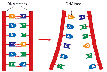
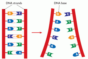
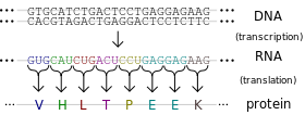
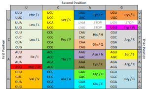
  • As I mentioned above, genes tell your cells how to make proteins. Genes are made of DNA. DNA is made of tiny building blocks called nucleotides. There are four kinds of DNA building blocks. These building blocks are grouped in bundles of three. Every one of those three DNA building block bundles tells the cell how to make a little piece of the protein. Those bundles are called codons. Then the three DNA building blocks next to that bundle tell the cell how to make another little piece of the protein, and so on. Those little pieces stick together and make the full sized protein.
  • The cell is able to make a protein by reading through this gene three pieces at a time. This is how our cells use genes to make proteins.
  • So now we know that those three DNA building blocks work together to me a tiny piece of protein. There are four kinds of DNA building blocks. What piece of protein is made is determined by which three building blocks are grouped together. There are many combinations of building blocks.
  • If one of those building blocks is mutated, it could cause the wrong protein piece to be made. So it seems that each of the three building blocks is really important to making the right protein. However, this is not the case. In fact, of those three building blocks, the last one is mostly irrelevant. If it’s mutated, it will usually still make the right protein piece. In some cases, the second one isn’t important either. The first one is the most important, but having a mutation there doesn’t always mean the protein is made wrong either.
  • So genes could potentially have up to 1/3 of its building blocks mutated without causing a problem. (I’m being very general here.) This phenomenon, in which the third DNA building block in a group can be wrong without messing up the protein is called wobble. Wobble is a built in mechanism that allows cells to make mutations sometimes without consequences.
  • That’s a lot of mutations that don’t cause problems. That’s a lot of mutations that still allow the cell to make the right protein. That’s a lot of mutations without causing symptoms or disease.
  • Your genes can withstand a lot of changes to single building blocks. When a single nucleotide is changed, it is called a single nucleotide polymorphism (snp). Most of these snps don’t cause trouble at all. The only way to know which ones cause problems is to gather up a bunch of people with these mutations and study them to see if they have diseases and, if so, which ones. So just having a mutation is not enough to know if it causes diseases without further study.

For additional reading, please visit the following posts:
Gene expression and the CKIT D816V mutation
The MastAttack 107: The Layperson’s Guide to Understanding Mast Cell Diseases, Part 2
The difference between C117+ and CKIT+
Heritable mutations in mastocytosis

The MastAttack 107: The Layperson’s Guide to Understanding Mast Cell Diseases, Part 2

I have answered the 107 questions I have been asked most in the last four years. No jargon. No terminology. Just answers.

3. What causes mast cell disease?

  • The cause of mast cell disease is not yet definitively known.
  • As mentioned yesterday, when the body makes too many copies of a broken cell, those cells are called ‘clonal’ cells. In clonal forms of mast cell disease, the bone marrow makes too many mast cells. Those mast cells also don’t work correctly. Examples of clonal mast cell diseases are systemic mastocytosis and cutaneous mastocytosis.
  • Patients with systemic mastocytosis often have a specific genetic mutation called the CKIT D816V mutation. About 80-90% of systemic mastocytosis patients have this mutation. This mutation is in mast cells and it tells the mast cells to stay alive WAY longer than they should. And mast cells already live for months or years, a very long time for cells to live in the body. So patients with this mutation can end up with way too many broken mast cells.
  • Despite the fact that we know that many patients have this mutation, we do not say that this mutation CAUSES the disease. The reason for this is that sometimes, mast cell patients don’t have the mutation when they get sick but they develop it later. Sometimes, mast cell patients have the mutation and then lose it later. So we are still looking for something that causes the disease.
  • Patients with non-clonal mast cell disease do not have a single major mutation like the CKIT D816V mutation. This makes it harder to diagnose. Researchers have found that many times, patients with MCAS DO have mutations similar to the ones systemic mastocytosis patients do. But the MCAS patients often have different mutations from each other. That’s why it’s not helpful yet for diagnosis.
  • Despite the fact that the mutations described here are not considered to be heritable, there is more and more evidence that mast cell disease can happen to many people in the same family. See the next question for more details.

4. Is mast cell disease heritable?

  • Mast cell disease often affects multiple members of the same family. Importantly, patients often have a different type of mast cell disease than their relatives. This implies that mast cell disease is more of a spectrum rather than several different diseases.
  • A survey found that 74% of mast cell patients interviewed reported at least one first degree relative that had mast cell disease. This same study found that 46% of those patients had mast cell disease that affected more than just their skin. This is called systemic disease.
  • The CKIT D816V mutation is the mutation most strongly associated with clonal mast cell disease. The CKIT D816V mutation is NOT heritable.
  • There are very rare instances of other heritable mutations in families that have mast cell disease. The significance of this is not clear.

5. Can mast cell disease be cured?

  • Generally speaking, there is no cure for mast cell disease.
  • Children who present with cutaneous mastocytosis sometimes grow out of their disease. Their lesions disappear. Their mast cell symptoms affecting the rest of the body may disappear. We do not know why this happens. It has been heavily researched with long term follow up of children with childhood mastocytosis (at least one paper followed them for 20 years).
  • Children with true systemic mastocytosis do not grow out of their disease.
  • There is not yet data on children with MCAS. Anecdotally, they do not seem to grow out of their disease like kids with cutaneous mastocytosis can. Importantly, this is just what it looks like to me. Again, there is no data.
  • People with adult onset mast cell disease have lifelong disease.
  • There is one notable exception to this scenario. There are reports of curing mast cell disease following hematopoietic stem cell transplant/bone marrow transplant.
  • Transplantation is EXTREMELY dangerous. The transplant is MUCH, MUCH more dangerous than mast cell disease. Many people do not survive the protocol necessary to prepare for transplant. Many die from complications, or from a disease they acquired after their transplant.
  • Rarely, people may have malignant forms of mast cell disease, aggressive systemic mastocytosis (ASM) or mast cell leukemia (MCL). A few patients with these diseases have tried transplants after everything else failed. While some did see improvement after transplant, no one has survived more than a few years.
  • Conversely, sometimes people with mast cell disease have these transplants for other reasons, like having another blood cancer or bone marrow disease that requires transplant. In this group of people, some see drastic improvement of their mast cell disease. Some see a full remission of mast cell disease. Some do not get any improvement. These findings are pretty recent so it’s hard to be more specific.

For more detailed reading, please visit these posts:

The Provider Primer Series: Introduction to Mast Cells

The Provider Primer Series: Mast cell activation syndrome (MCAS)

The Provider Primer Series: Cutaneous Mastocytosis/ Mastocytosis in the Skin

The Provider Primer Series: Diagnosis and natural history of systemic mastocytosis (ISM, SSM, ASM)

The Provider Primer Series: Diagnosis and natural history of systemic mastocytosis (SM-AHD, MCL, MCS)

Mast cell disease in families

Heritable mutations in mastocytosis

Take home points: October 2015

Childhood mastocytosis: Update

  • Cutaneous mastocytosis in children is the most common form of mastocytosis
  • True systemic mastocytosis is very rare in children
  • An NIH study of 105 children found 30-65% improved over time
  • Elevated baseline tryptase level and organ swelling were good indicators of SM
  • Serum tryptase should be measured every 6-12 months
  • Children with swelling of both liver and spleen were positive for CKIT D816V mutation
  • Swelling of liver and spleen together was linked to disease persisting into adulthood
  • Most children with UP with skin and minor GI issues had normal tryptase
  • Diffuse cutaneous mastocytosis patients had a much higher average tryptase but no organ swelling
  • Serum tryptase and IgE were inversely related (high tryptase with low IgE, low tryptase with high IgE)

Chronic mast cell leukemia: a new variant of systemic mastocytosis

  • Mast cell leukemia (MCL) has a significantly shortened lifespan
  • Usually over 20% of nucleated cells in bone marrow are atypical mast cells
  • Mast cells are present in large quantities on the blood
  • Cases where less than 10% of white blood cells in blood are mast cells are called aleukemic variant MCL
  • Cases where over 20% of nucleated cells in bone marrow are mature mast cells are called chronic MCL
  • Chronic MCL patients do not have any C findings (the clinical markers for SM patients associated with very aggressive disease)
  • Chronic MCL patients have stable disease state but can progress to acute MCL at any time
  • Mediator release symptoms are more common in chronic MCL than acute MCL
  • Acute MCL is marked by immature CD25+ mast cells
  • Acute MCL patients do have C findings (the clinical markers for SM patients associated with very aggressive disease)
  • Acute MCL has a very short survival time, usually less than a year

The effects of cortisol on mast cells: Part 2 of 3

Glucocorticoids, like cortisol, can affect mast cells in many ways. As I discussed in my previous post, there are many ways for mast cells to release mediators when activated. In all of these pathways, there are many molecules involved that carry the signal, like people passing the Olympic torch. In mast cells, one of the molecules that suppresses inflammatory activation signal is called SLAP (yes, really).  Cortisol increases the amount of SLAP in mast cells so inflammatory activation signals are suppressed.

An important step in degranulation is changing the amount of calcium inside the cell and moving it to different parts of the cell. In some studies, glucocorticoids can affect this movement of calcium. Other studies have found that in some pathways, glucocorticoids don’t affect calcium movement, but instead interfere with things like the IgE receptor.

Cortisol is also thought to directly inhibit stem cell factor (SCF) binding to the CKIT receptor. When SCF binds to the CKIT receptor, this sends a signal to the mast cell to stay live.  This means that taking glucocorticoids can let mast cells die at the appropriate time. SCF also tells mast cells to go to inflamed spaces.  By blocking this signal, glucocorticoids suppress inflammation.

One of the ways that molecules carry a signal is by changing the next molecule in the pathway. A big way that cells changing molecules is by chopping off a piece of them called a phosphate group.  This is done by special enzymes called phosphatases.  Glucocorticoids affect the availability of phosphatases so they aren’t able to get to the right part of the cell to carry the signal.  When this happens, there is less activation and less histamine release.

Arachidonic acid is the molecule modified to make eicosanoids (leukotrienes, thromboxanes and prostaglandins.) Glucocorticoids directly interfere with the production of these molecules in multiple ways.  The first way is by interfering with COX-2, one of the enzymes that makes prostaglandins.  Another way is by preventing arachidonic acid from being released to a place where they can be turned into leukotrienes, thromboxanes and prostaglandins.  This occurs because glucocorticoids increase the amount of a powerful anti-inflammatory molecule called annexin-I.  Annexin-I inhibits the molecule that releases the arachidonic acid, called phospholipase A2.

Annexin-I was the subject of an important paper earlier this year. In trying to identify exactly how mast cell stabilizers like ketotifen and cromolyn work, the researchers discovered that treatment with mast cell stabilizers decreased degranulation and increased annexin-I made by mast cells.  They also found that glucocorticoids had the same effect.

References:

Oppong E, et al. Molecular mechanisms of glucocorticoid action in mast cells. Molecular and Cellular Endocrinology 2013: 380, 119-126.

Varghese R, et al. Association among stress, hypocortisolism, systemic inflammation and disease severity in chronic urticaria. Ann Allergy Asthma Immunol 2016: 116, 344-348.

Zappia CD, et al. Effects of histamine H1 receptor signaling on glucocorticoid receptor activity. Role of canonical and non-canonical pathways. Scientific Reports 2015: 5.

Coutinho AE, Chapman KE. The anti-inflammatory and immunosuppressive effects of glucocorticoids, recent developments and mechanistic insights. Mol Cell Endocrinol 2011: 335(1), 2-13.

Sinniah A, et al. The role of the Annexin-A1/FPR2 system in the regulation of mast cell degranulation provoked by compound 48/80 and in the inhibitory action of nedocromil. International Immunopharmacology 2016: 32, 87-95.

The difference between CD117+ and CKIT+

Hey, everyone –

I received a request to clarify the difference between being CD117+ and CKIT+.

CD117 is a receptor on the outside of mast cells. It is normal and all mast cells are CD117+. This is how we identify them as mast cells. If you have a bone marrow biopsy done and it says no CD117 is found, this is not because there are no mast cells there. It is because the test for CD117 isn’t sensitive enough to find those few mast cells. This is called the limit of detection (LoD).

When there is more of something present, it is easier to find it. Say I am in a field and there are five tennis balls scattered. If I walk around for a long time, maybe I will find three tennis balls. But if there is only one tennis ball to be found, I may not find it. I have less of a chance of finding it because there aren’t as many so it’s harder.

Being CD117+ is NORMAL for mast cells. It just means that it’s a mast cell. But mast cells that are constantly activated have more CD117+ on their outside membranes. Think of it like the tennis balls – if there are five CD117 receptors on a mast cell, it’s easier for the test to find one. If there is only one, the test might miss it.

CD117 is also called the CKIT receptor. It is a receptor that gives mast cells the signal to stay alive and encourage more mast cells to mature. If you get a biopsy report back and it is CD117+, then it will say CD117. The reason the report doesn’t call it positive for CKIT is historical and has to do with the fact that it was identified first as CD117 and later called CKIT because of similarities with other proteins of similar names.

When mast cell patients say CKIT+, it is a misnomer. It means that they are positive for the D816V mutation in CKIT, which is a marker for systemic mastocytosis. So being CD117+ and CKIT+ are not the same. CD117+ just means mast cell. CKIT+ (D816V) means neoplastic mast cell.

The D816V mutation changes the shape of the CD117 (CKIT) receptor and tells the mast cell to stay alive and encourage other mast cells to mature even when it shouldn’t.

Being CD117+ does not affect medication profile for mast cell disease at all. It just means it’s a mast cell. Some drugs are approved only for CKIT- patients (negative for D816V).

CD117/CKIT is a tyrosine kinase, which is a kind of protein. There are hundreds of known tyrosine kinases, CD117/CKIT is just one. Tyrosine kinase inhibitors can affect cells by blocking the signal to stay alive. Tyrosine kinases do not take up tyrosine from the environment, it has literally nothing to do with tyrosine metabolism at all.

If there any questions, ask in the comments.

What do all these words mean? (Part 2)

What does it mean if a person is CD117 positive in a biopsy? Is this bad?

In the context of mast cell disease, it usually just means that mast cells were found.


If CD117 is normal for mast cells, why are people sometimes “negative for CD117” on biopsies?

This sometimes happens. When you have mast cell disease, you often have more CD117 receptors on mast cells. This makes it easier for the test to find them. So when you are found to be “negative for CD117” in regards to mast cell disease, you are not truly “negative”. You just express a lower amount of CD117 receptors so the test didn’t see them.


I am a mast cell patient and my bone marrow biopsy was positive for CD117. How do I know that it is normal mast cells that show CD117 and not these other dangerous cells you mentioned?

You can tell by looking at the cells with special stains. Pathologists and immunohistochemistry scientists are very skilled at distinguishing one cell type from another. They can tell based upon what the cell looks like in addition to being positive for CD117.


If I am CD117 positive in a biopsy, does that mean I am “CKIT positive”?

No. If I could do away with one single phrase in mast cell terminology, it would be CKIT+.

CKIT+ is a term used to mean “positive for the D816V mutation in codon 816 of the CKIT gene”. It means you are positive for a mutation that has been associated with neoplastic disorders of mast cells. So when people say they are CKIT+, they mean they were found to have a mutation. They do NOT mean they were found to have CKIT/CD117 on their mast cell surfaces, because this is totally normal and is the case for everyone.

Additionally, the test to detect CD117 on a cell surface is NOT the same test used to identify the D816V mutation. That test breaks open the cells and looks for a specific mutation in the DNA sequence. They are not run at the same time.


Why is it important to know if I am positive for the D816V mutation (CKIT+)?

The D816V mutation changes the shape of the CKIT receptor. Due to this wrong shape, the receptor does not need SCF to bind to the receptor to tell the mast cell to live longer. In this new shape, the receptor is stuck in an “activated” position, so it is telling the cell to live longer all the time, without SCF. This is called “autoactivation”.

The D816V mutation is one of the minor criteria for systemic mastocytosis, so it is important for classification purposes. Also, it may affect your treatment plan in the unlikely situation of needing chemotherapy.


What is CD25?

CD25 is part of a receptor for a molecule called IL-2. Normally, mast cells do not express receptors for IL-2, which is a molecule that regulates development of T cells. When mast cells express CD25, it is an indication that the mast cell is neoplastic. Many T cells normally express CD25, so if it is on a biopsy report, keep in mind that it’s abnormal on mast cells, but not everywhere.

Presence of CD25 on mast cells is one of the minor criteria for SM.


What is CD2?

CD2 is an example of a “CD” molecule that is not a receptor. It is a cell adhesion molecule so it helps cells stick together. Normally, mast cells do not express CD2. When mast cells express CD2, it is an indication that the mast cell is neoplastic. Many T cells normally express CD25, so if it is on a biopsy report, keep in mind that it’s abnormal on mast cells, but not everywhere. CD2 is a less accurate indication of SM than CD25.

Presence of CD2 on mast cells is one of the minor criteria for SM.


What is CD30?

CD30 is a receptor for proteins associated with tumor necrosis factor. It is commonly referred to as a tumor marker, but this is not always the case. CD30 has recently been shown to be frequently positive in patients with all forms of SM (ISM, SSM, SM-AHNMD, ASM). However, on other cells besides mast cells, it may indicate lymphoma or other conditions.


What is CD34?

CD34 is another cell adhesion molecule. It is thought to allow stem cells to attach to proteins in the bone marrow. It is found on many progenitor cells, cells that later become other kinds of cells. Mast cells express CD34, though this tends to be lost as they move into tissues.

What do all these words mean? (Part 1)

What is a neoplasm?

A neoplasm is an abnormal growth of cells. Systemic mastocytosis is a neoplasm because your body makes too many mast cells. Cancers are all neoplasms, but not all neoplasms are cancers. SM is not cancer.


What is a myeloproliferative neoplasm (MPN)?

Myelo- means “related to granulocytes”, cells that store chemicals in granules. Mast cells are a type of granulocyte.

Proliferative means “cell growth”.

So together you have “growth of too many granulocytes”.


What are receptors?

Receptors are proteins on the outside of cells. They have very unique and specific shapes, but it is easier to think of them as being shaped by cups. Only very specific molecules fit into these cups. When the molecule is in the receptor, the cell knows to do something. What this something is varies a lot from receptor to receptor.

For example, when an IgE antibody binds to the IgE receptor on a mast cell, the mast cell degranulates. However, not any molecule would be able to bind in the IgE receptor and cause this action.


What are antibodies?

These are large proteins that help the immune system identify and destroy things like bacteria and viruses. Sometimes your body mistakenly makes antibodies to things inside the body. This causes autoimmune disease.

In labs, antibodies are very useful. There are ways to make antibodies to almost anything in the lab. Using these lab made antibodies, scientists are able to test for specific structures that tell us what cells are present in a sample and how the cells are working.


What are immunoglobulins?

They are the same as antibodies.


What does CD mean? Like in CD117?

CD means “cluster of differentiation”. This means that it is a protein or group of proteins on the surface of a cell that is recognized by an antibody. This means that in a lab, if I use an antibody called “ABC123” and it binds to a specific protein on the outside of cells, that protein will be called “CD-ABC123”.

Over time, as we learn more about “CD-ABC123”, we may realize that this protein is made by a gene called “Wow”. So some people will call it “Wow” and some will continue to call it “CD-ABC123”, which can be confusing. Generally speaking, scientists who work with antibody testing usually use the “CD-ABC123” name and doctors use the “Wow” name. However, both names are still correct.

Receptors are often given “CD” names, but not all “CD” molecules are receptors. Some “CD” molecules are on the outside of cells to do other things, like help cells stick to other cells.


What is CKIT?

CKIT is a receptor that is found on all mast cells, whether or not a person has mast cell disease. The CKIT receptor is also called CD117. They are the same thing. CKIT is sometimes also called KIT. They are also the same thing.

The molecule that fits in the CKIT receptor is called stem cell factor (SCF). When SCF binds the CKIT receptor, it tells the mast cell to stay alive much longer than it is supposed to. It also starts a process that tells mast cells to make more mast cells.


CKIT (CD117) is only found on mast cells?

No, other normal cells have CKIT (CD117) on their surface. Epithelial cells in the skin, breast and some parts of the brain express CD117. Some stem cells in the bone marrow and melanocytes also express CD117. Smooth muscle and fibroblasts do not express CD117. This is important because smooth muscle and fibroblasts are often found close to the cells we might be looking for that may be positive for CD117.

However, when found on cells that don’t normally express CD117, it can be a sign of cancer – but ONLY if these cell types don’t normally have CD117 on their surfaces. Examples of cancers that express CD117 include angiosarcoma and Ewing sarcoma.

Mast cell mutations: TET2 and mutation profiles of aggressive subtypes

TET2 (Tet methylcytosine dioxygenase 2) is found to be mutated in 20.8-29% of SM patients. Of note, dozens of mutations have been identified in this gene, including missense, nonsense, frameshift and deletion mutations. These mutations cause formation of a defective and less active TET2 enzyme. TET2 is located at chromosome 4q24 and mutations at this location are associated in both MPN and MDS conditions.

TET2 is involved in DNA methylation and demethylation, although the exact nature of this involvement is not clear. When a methyl group is added to a cytosine at a specific place in front of a gene, the gene is turned off and is not expressed. This is called “methylation.” TET2 adds a hydroxyl group to 5-methylcytosine, but it is not well understood if this turns the gene off. TET2 may also be involved in demethylating DNA, or removing those specific methyl groups. It has been shown to be involved with DNA demethylation during bone development.

One study looked at the mutational profiles of patients with various forms of SM, including ISM, SSM, SM-AHNMD, ASM and MCL, all of whom were positive for CKIT D816V mutation. 15/39 had a TET2 mutation. None of those patients had ISM or SSM. Of those with an aggressive form and a TET2 mutation, 67% had more than one TET2 mutation.

In this study, 24/27 patients with advanced SM (SM-AHNMD, ASM, MCL) had mutations beyond the D816V mutation. 5/5 SM-AHNMD patients and 19/22 ASM or MCL patients had multiple mutations (CKIT and something else.) In contrast, only 3/12 ISM or SSM patients had additional mutations. In advanced SM, 78% had at least 3 mutations, and 41% had at least 5.

These mutational profiles have clear implications clinically. 96% patients with major blood abnormalities (anemia <10 g/dL and/or thrombocytopenia < 100 x 10e9/L in addition to monocytosis > 1 x 10e9/L and/or eosinophilia >10%) had at least one additional molecular mutation regardless of SM subtype.

Advanced SM patients in this study all had one of the following multiple mutation profiles: 26% KIT-TET2-SRSF2, 18% KIT-SRSF2-RUNX1, 13% KIT-TET2-CBL, 10% KIT-SRSF2-ASXL1 10%, and 10% KIT-TET2-ASXL1. Patients with advanced SM (and therefore multiple mutations) were also found to be significantly older (68 years of age on average) than those with just the CKIT mutation (48 years of age on average.)

Having a TET2 mutation seems to predispose myeloid cells to become neoplastic later in life. It is important to distinguish that the TET2 mutation seems to “allow” this transformation rather than causing it. In mice who don’t have the TET2 gene and thus don’t have the TET2 enzyme, stem and progenitor cells have trouble maintaining balance and spontaneously become neoplastic later in life. In TET2 deficient cells, mast cells with D816V mutation show increase in proliferation and survival as opposed to those without with normal TET2 levels. Presence of TET2 in addition to the presence of CKIT D816V mutation is associated with more aggressive forms of SM (including ASM, MCL and SM-AHNMD.)


References:

Damaj, G., Joris, M., Chandersris, O., Hanssens, K., Soucie, E., Canioni, D., et al., 2014.ASXL1 but not TET2 Mutations Adversely Impact Overall Survival of PatientsSuffering Systemic Mastocytosis with Associated Clonal Hematologic Non-Mast-Cell Diseases. PLoS ONE 9 (1), e85362.

Schwaab, J., Schnittger, S., Sotlar, K., Walz, C., Fabarius, A., Pfirrmann, M., et al., 2013.Comprehensive mutational profiling in advanced systemic mastocytosis. Blood122 (October (14)), 2460–2466.

Soucie, E., Hanssens, K., Mercher, T., Georgin-Lavialle, S., Damaj, G., Livideanu, C.,et al., 2012. In aggressive forms of mastocytosis. TET2 loss cooperates with c-KITD816V to transform mast cells. Blood 120 (December (24)), 4846–4849.

Soucie, E., Brenet, F., Dubreuil, P. Molecular basis of mast cell disease. Molecular Immunology 63 (2015) 55-60.

Heritable mutations in mastocytosis

While the most well-known mutation associated with SM is the CKIT D816V, there are numerous other mutations that can contribute to mast cell disease and presentation. The CKIT gene produces a tyrosine kinase receptor on the outside of the mast cell. Tyrosine kinases function as switches that turn certain cell functions on and off. When stem cell factor binds to the CKIT receptor, it turns on the signal for the mast cell to live longer than usual and to make more mast cells.

The D816V mutation is located in a specific part of the CKIT gene called exon 17. As many as 44% of SM patients have CKIT mutations outside of exon 17, either alone or in addition to the D816V mutation. (Please note that for the purposes of this post, SM is used to refer to SM, ASM and SM-AHNMD in keeping with the source literature.) Still, most doctors and researchers believe the D816V mutation is not heritable. This has important implications because it means many doctors also believe mast cell disease is sporadic and not heritable.

Almost 75% of MCAD (SM, MCAS, MCL) patients had at least one first degree relative with MCAD. This study, published in 2013, demonstrated that despite the non-heritable nature of the D816V mutation, mast cell disease is indeed heritable. Currently, four heritable mutations present in mast cell patients have been identified.

CKIT is often called KIT. In one family in which the mother, daughter and granddaughter have all have indolent SM, they were all found to have a deletion at position 409 in KIT (called KITdel409.) The KIT F522C mutation has been associated with ISM.   Another heritable mutation, KIT K509I, has been identified multiple times by different researchers. The first publication to identify this mutation was published in 2006. It has been found in a mother/daughter set who have ISM, and in another mother/daughter set in which the mother has ASM and the daughter has CM. This mutation was noted in a 2014 paper to be associated with well differentiated SM.

There have been reports of families in which multiple members with ISM or SM-AHNMD had the D816V mutation. Importantly, in these patients, the mutation was readily found in numerous cell types, including mast cells, CD34+ hematopoietic precursor cells, blood leukocytes, oral epithelial cells, blast cells and erythroid precursors. Despite this finding, the majority of literature continues to report the D816V mutation as not heritable.


References:

G.J. Molderings. The genetic basis of mast cell activation disease – looking through a glass darkly. Critical Reviews in Oncology/Hematology 2014.

G.J. Molderings, B. Haenisch, M. Bogdanow, R. Fimmers, M.M. Nöthen. Familial occurrence of systemic mast cell activation disease. PLoS One, 8 (2013), p. e76241

Hartmann, E. Wardelmann, Y. Ma, S. Merkelbach-Bruse, L.M. Preussenr, C. Woolery, et al. Novel germline mutation of KIT associated with familial gastrointestinal stromal tumors and mastocytosis. Gastroenterology, 129 (2005), pp. 1042–1046

R.A. Speight, A. Nicolle, S.J. Needham, M.W. Verrill, J. Bryon, S. Panter. Rare germline mutation of KIT with imatinib-resistant multiple GI stromal tumors and mastocytosis. J Clin Oncol, 31 (2013), pp. e245–e247

de Melo Campos, J.A. Machado-Neto, A.S.S. Duarte, R. Scopim-Ribeiro, F.F. de Carvalho Barra, J.Vassallo, et al.Familial mastocytosis: identification of KIT K509I mutation and its in vitro sensitivity to imatinib, dasatinib and PK412. Blood, 122 (2013), p. 5267

L.Y. Zhang, M.L. Smith, B. Schultheis, J. Fitzgibbon, T.A. Lister, J.V. Melo, et al. A novel K5091 mutation of KIT identified in familial mastocytosis – in vitro and in vivo responsiveness to imatinib therapy. Leukemia Res, 30 (2006), pp. 373–378

E.C. Chan, Y. Bai, A.S. Kirshenbaum, E.R. Fischer, O. Simakova, G. Bandara, et al. Mastocytosis associated with a rare germline KIT K509I mutation displays a well-differentiated mast cell phenotype. J Allergy Clin Immunol, 134 (2014), pp. 178–187

Akin, G. Fumo, A.S. Yavuz, P.E. Lipsky, L. Neckers, D.D. Metcalfe. A novel form of mastocytosis associated with a transmembrane c- Kit mutation and response to imatinib. Blood, 103 (2004), pp. 3222–3225

Escribano, R. Nunez-Lopez, M. Jara, A. Garcia-Montero, A. Prados, C. Teodosio, et al. Indolent systemic mastocytosis with germline D816 V somatic c-kit mutation evolving to an acute myeloid leukemia. J Allery Clin Immunol, 117 (Suppl.) (2006), p. S125

Gene expression and the D816V mutation

What exactly is the D816V mutation and why does it matter?  To answer that, we need to understand the basic pathway by which a cell expresses a gene.    

DNA (deoxyribonucleic acid) is the molecule that contains the genetic code for all known living organisms and some viruses.  DNA is composed of two strands that wrap around each other in a double helix pattern.  DNA is built out of nucleotides, molecules that contain energy.  The nucleotides that build DNA are adenine (A), guanine (G), thymine (T) and cytosine (C).  These nucleotides bond in specific pairs.  This means that when one nucleotide in on one strand of DNA, there is a specific nucleotide on the other strand.  A and T, and C and G specifically bond to each other.  They are known as base pairs.  DNA strands made up of base pairs are said to be “complementary.” 


RNA (ribonucleic acid) is a more versatile nucleic acid that codes, regulates and expresses genes, amongst other things.  It also has base pairs: adenine and uracil (U), and thymine and cytosine.  These nucleotides can be complementary to DNA nucleotides.  For example, an RNA adenine is complementary to a DNA thymine, and so on.

DNA replication is the process by which an exact copy of a piece of DNA is made.  This happens when a cell divides.  In replication, the DNA double helix “unzips,” or splits apart into two strands, the base pairs of which are not connected.  Special enzymes move along each of the two split strands and place the appropriate nucleotides next to each strand to form base pairs.  The end result of this is two double helices of DNA that are exact copies.   


Some parts of DNA, called genes, tell the cell how to make proteins or RNA that has a specific function.  (Sometimes RNA can also do this.)  Genes tell the cell how to build and maintain the cell and allow it pass on traits to offspring.  These proteins or RNA are made by expressing the gene.  In gene expression, the information from the gene is turned into a “gene product,” that will be made into something useful for the cell.
Transcription is the start of gene expression.  Gene expression is very complicated and controlled by many mechanisms.  Having a gene does not mean it will always be expressed.  In transcription, a piece of DNA is copied into a complementary RNA strand.   This RNA is called messenger RNA (mRNA.)  This is a complicated process with several steps.  Once a gene is translated, the mRNA with the gene code goes to the ribosome, a place in the cell that makes proteins.  Proteins are made of amino acids. 

So how exactly does the DNA code for the protein the ribosome will make? Let’s focus on that.
The ribosome reads the messenger RNA made from the DNA gene three nucleotides at a time. Again, when using the code to build a protein, the ribosome reads the code in blocks of three nucleotides. These blocks of three nucleotides are called “codons.” Every combination of three-nucleotides tells the ribosome to add a specific amino acid to the protein. The majority of genes are encoded using this same codon code. So by knowing the DNA sequence, we can anticipate the amino acids that build the protein encoded by the gene. 


How does the ribosome know where to start?  There’s a start codon.  (And some other things also.)
How does the ribosome know where to stop?  There’s a stop codon.  (And some other things also.)
There are several types of genetic mutations, or alterations of the code from the one seen in most of the population.  In a point mutation, a single nucleotide is changed.  The D816V mutation is a point mutation. 
We use a specific nomenclature to describe genetic mutations.  Amino acids are often referred to with single letter codes for the sake of brevity.  The amino acid aspartic acid is referred to as “D,” while the amino acid for valine is referred to as “V.”  In the CKIT gene, the amino acid sequence Asp-Phe-Gly (aspartic acid – phenylalanine – glycine) is very important to the receptor being shaped the right way. 
Aspartic acid is encoded by the RNA code “GAU” or “GAC.”  In cells with the D816V mutation, this sequence is changed to “GUU” or “GUC.”  The second base is changed from an A to a U.  Doing this changes the amino acid encoded from aspartic acid (D) to valine (V).  These amino acids are shaped differently, and because of this, the receptor is shaped differently and behaves differently.  When the receptor is made with the amino acid aspartic acid in that place, SCF (stem cell factor) binds to the receptor and activates the cell, telling it not to die and to make more cells.  When the receptor is made with the amino acid valine in that place, the receptor activates itself and SCF is not needed.  It basically tells itself not to die and to make more cells repeatedly. 
So the term “D816V” means that at codon 816, the code was altered in a way that changed the amino acid for aspartic acid to valine.  Some people with mast cell disease don’t have the D816V mutation, but often they have another mutation at codon 816, like D816G.  Sometimes they have a mutation somewhere else in the same “exon.”  An exon is the part of the code that is sent as RNA to be made into a gene product.  The location of the CKIT gene is referred to as exon 17.  

Image credits:


http://www.brooklyn.cuny.edu/bc/ahp/BioInfo/TT/TscriptD.html

http://genmed.yolasite.com

http://www.bristol.k12.ct.us/

http://en.wikipedia.org/wiki/Gene_expression