Skip to content

methylation

Mast cell mutations: TET2 and mutation profiles of aggressive subtypes

TET2 (Tet methylcytosine dioxygenase 2) is found to be mutated in 20.8-29% of SM patients. Of note, dozens of mutations have been identified in this gene, including missense, nonsense, frameshift and deletion mutations. These mutations cause formation of a defective and less active TET2 enzyme. TET2 is located at chromosome 4q24 and mutations at this location are associated in both MPN and MDS conditions.

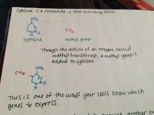
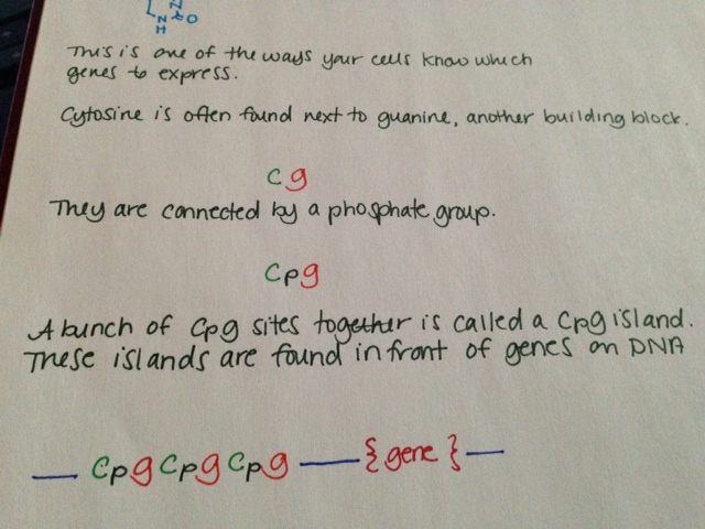
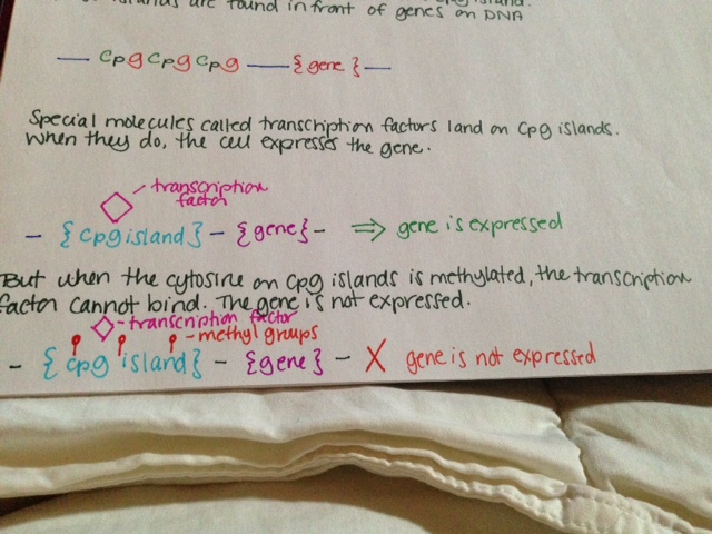
TET2 is involved in DNA methylation and demethylation, although the exact nature of this involvement is not clear. When a methyl group is added to a cytosine at a specific place in front of a gene, the gene is turned off and is not expressed. This is called “methylation.” TET2 adds a hydroxyl group to 5-methylcytosine, but it is not well understood if this turns the gene off. TET2 may also be involved in demethylating DNA, or removing those specific methyl groups. It has been shown to be involved with DNA demethylation during bone development.

One study looked at the mutational profiles of patients with various forms of SM, including ISM, SSM, SM-AHNMD, ASM and MCL, all of whom were positive for CKIT D816V mutation. 15/39 had a TET2 mutation. None of those patients had ISM or SSM. Of those with an aggressive form and a TET2 mutation, 67% had more than one TET2 mutation.

In this study, 24/27 patients with advanced SM (SM-AHNMD, ASM, MCL) had mutations beyond the D816V mutation. 5/5 SM-AHNMD patients and 19/22 ASM or MCL patients had multiple mutations (CKIT and something else.) In contrast, only 3/12 ISM or SSM patients had additional mutations. In advanced SM, 78% had at least 3 mutations, and 41% had at least 5.

These mutational profiles have clear implications clinically. 96% patients with major blood abnormalities (anemia <10 g/dL and/or thrombocytopenia < 100 x 10e9/L in addition to monocytosis > 1 x 10e9/L and/or eosinophilia >10%) had at least one additional molecular mutation regardless of SM subtype.

Advanced SM patients in this study all had one of the following multiple mutation profiles: 26% KIT-TET2-SRSF2, 18% KIT-SRSF2-RUNX1, 13% KIT-TET2-CBL, 10% KIT-SRSF2-ASXL1 10%, and 10% KIT-TET2-ASXL1. Patients with advanced SM (and therefore multiple mutations) were also found to be significantly older (68 years of age on average) than those with just the CKIT mutation (48 years of age on average.)

Having a TET2 mutation seems to predispose myeloid cells to become neoplastic later in life. It is important to distinguish that the TET2 mutation seems to “allow” this transformation rather than causing it. In mice who don’t have the TET2 gene and thus don’t have the TET2 enzyme, stem and progenitor cells have trouble maintaining balance and spontaneously become neoplastic later in life. In TET2 deficient cells, mast cells with D816V mutation show increase in proliferation and survival as opposed to those without with normal TET2 levels. Presence of TET2 in addition to the presence of CKIT D816V mutation is associated with more aggressive forms of SM (including ASM, MCL and SM-AHNMD.)


References:

Damaj, G., Joris, M., Chandersris, O., Hanssens, K., Soucie, E., Canioni, D., et al., 2014.ASXL1 but not TET2 Mutations Adversely Impact Overall Survival of PatientsSuffering Systemic Mastocytosis with Associated Clonal Hematologic Non-Mast-Cell Diseases. PLoS ONE 9 (1), e85362.

Schwaab, J., Schnittger, S., Sotlar, K., Walz, C., Fabarius, A., Pfirrmann, M., et al., 2013.Comprehensive mutational profiling in advanced systemic mastocytosis. Blood122 (October (14)), 2460–2466.

Soucie, E., Hanssens, K., Mercher, T., Georgin-Lavialle, S., Damaj, G., Livideanu, C.,et al., 2012. In aggressive forms of mastocytosis. TET2 loss cooperates with c-KITD816V to transform mast cells. Blood 120 (December (24)), 4846–4849.

Soucie, E., Brenet, F., Dubreuil, P. Molecular basis of mast cell disease. Molecular Immunology 63 (2015) 55-60.

DNA Methylation: How it works

DNA methylation is one of the ways your cells control which genes to express. It is an example of epigenetic modification. Epigenetics mechanisms like this do not change the DNA sequence, only the way the genes are expressed. Whether or not DNA methylation is heritable is not clear.

This is how DNA methylation works:

  • Cytosine is a nucleotide, a DNA building block.
  • Through the action of an enzyme called methyltransferase, a methyl group is added to cytosine.
  • This is one of the ways your cells know which genes to express.
  • Cytosine is often found next to guanine, another building block. This is sometimes shown in literature as “CG.”
  • Cytosine and guanine are connected by a phosphate group. This is sometimes shown in literature as “CpG.”
  • A bunch of CpG sites together is called a CpG island. These islands are found in front of genes on DNA.
  • Special molecules called transcription factors land on CpG islands. When they do, the cell expresses the gene.
  • But when the cytosine on CpG islands is methylated, the transcription factor cannot bind. The gene is not expressed.

See pictures below.

Methylation is known to have an important role in cancer biology. Methylation of tumor suppressor genes causes the tumor suppressors not to be expressed, resulting in cancer.

Methylation 1Methylation 2Methylation 3

Genetics of MCAS: mutations and methylation

Despite having largely the same symptom profile as SM patients, people with MCAS often lack the signature D816V mutation, considered by many to be a marker of clonality and thus proliferation.  Despite the fact that their mast cells may look normal under a microscope, there is now a growing body of evidence indicating that mast cells in MCAS patients behave aberrantly due to mutations aside from D816V. 
In three studies that look at KIT mutations in MCAS patients, they were found in 26.5%, 44% and 65% of patients, respectively.  Even the average of these three values represents a significant number of people with MCAS who have KIT mutations.  Of note, these mutations are mostly outside of exon 17, where the D816V mutation is found.  In one patient, a mutation was found in the NLRP3 gene, associated with the inflammatory response. 
There are a number of other mutations in genes with various functions commonly found in patients with SM.  These include genes that make proteins to regulate other genes and genes that affect how we make proteins (epigenetic regulatory proteins, splicing machinery and transcription factors.)  To date, there have been no studies looking at whether MCAS patients have similar mutations.  However, there are clear hints that they do. 
The mutations previously mentioned were all somatic and not germline.  This means that the mutations arose after early embryonic development and thus were not heritable.  This fact indicates clearly that there are other germline mutations not yet identified that may induce the subsequent mutations.  This has been bolstered by a 2013 paper that found familial clustering in MCAS patients.   
An important finding is that nearly all mutations found in MCAS patients are heterozygous, meaning only one of the two copies was mutated.  This implies that homozygous mutations, in which both copies are mutated, could cause the cell to die.  Alternatively, the various mutations may work together to make the mutations work as strongly as if there were two mutated copies. 
At CpG sites, the cell can add a special marker to the cytosine called a methyl group.  This is called methylation.  If the cytosine in a gene is methylated, it turns the gene off.  When a gene is turned off, your body will not use that gene or make a protein from it.  This is one type of gene regulation (epigenetics.) 
Looking at the methylation status in the genomic DNA of white blood cells from MCAS patients shows aberrant methylation patterns.  The genes incorrectly methylated included some involved in DNA/RNA repair, DNA/RNA processing, cell death, cell activity and communication with other cells.  195 individual CpG sites have been identified as candidates as markers for MCAS.  Importantly, there is a correlation between the age of symptom onset and the year of birth, which indicates a sort of anticipation of developing MCAS.  This means that gene regulation by methylation could affect acquisition of later, non-heritable mutations like the ones seen in KIT.
A gene is made up of introns and exons.  When an RNA code is made from a gene to tell the cell how to make a protein, the cell cuts out some pieces of that RNA.  These pieces are called introns.  The remaining pieces, which are connected back together, are called exons.  The way the cell cuts the RNA and reconnects the pieces is called splicing.   The mutations in KIT seen in MCAS patients almost all involve intron and exon junctions, where they meet.  There are also some differences in the way splicing occurs in MCAS patients. 

Reference:
Molderings, Gerhard J.  The genetic basis of mast cell activation disease – looking through a glass darkly.  2014.  Critical Reviews in Oncology/Hematology.