Skip to content

MastAttack 107

The MastAttack 107: The Layperson’s Guide to Understanding Mast Cell Diseases, Part 73

86. What is the role of the spleen in systemic mastocytosis? (Part Two)

  • The spleen is basically a big filter for the blood. In the previous post, I mentioned one of its functions: to catch certain types of infections in the blood that your immune system has a hard time fighting in other ways.  It does some other things, too. The spleen stores red blood cells and platelets so that your body has a backup supply in case of hemorrhage or trauma.
  • The spleen also looks for something else when it filters the blood: damaged or abnormal blood cells. Damaged or abnormal blood cells get caught in the spleen so that they don’t continue to circulate in the blood. The spleen then breaks down those bad cells and uses materials from them to help make new healthy cells.
  • If there are lots of abnormal cells, then the spleen gets swollen because it is holding many more cells than usual. This is why the spleen swells in diseases where the body has abnormal cells in the blood stream. How much the spleen swells is directly proportional to the amount of abnormal cells in the blood.
  • For example, in acute leukemias, there are tons of abnormal cells circulating in the bloodstream. The spleen catches as many as they can. Because there are a lot, the spleen swells very quickly. In chronic leukemias, there are still abnormal cells, but they are produced at a much slower rate over time. This means that the spleen has more time to break down the broken blood cells it catches before it catches more of them. In these scenarios, the spleen swells more slowly over a longer period of time.
  • You can apply this understanding directly to mastocytosis. Patients with indolent systemic mastocytosis have fewer mast cells than those with smoldering or aggressive systemic mastocytosis, or mast cell leukemia. The patients with indolent systemic mastocytosis make some abnormal mast cells. The spleen will catch the ones it sees and remove them from the bloodstream. But mast cells don’t live in the blood and they only pass through the bloodstream for a short time. So the spleen has time to break down some mast cells before it catches more.
  • When a patient with indolent systemic mastocytosis starts to produce higher numbers of mast cells, that’s when you see the spleen starting to swell. That’s why spleen swelling is a B finding for systemic mastocytosis – it is an indicator that the body is making more mast cells than before, and could be headed toward a more aggressive form.
  • The number getting filtered out by the spleen increases so the spleen swells. The more abnormal mast cells produced, the more the spleen swells.
  • Additionally, when the bone marrow is making lots of aberrant mast cells, they are introduced into the blood stream in much larger numbers than normal. This means that they are more likely to get caught in the spleen than in a person with indolent systemic mastocytosis.
  • In smoldering systemic mastocytosis, the body makes more mast cells than in indolent systemic mastocytosis, so it’s more common for the spleen to swell. In aggressive systemic mastocytosis, the bone marrow is producing a lot of mast cells and many of them are caught in the spleen over a short period of time. In mast cell leukemia, even more are made and caught, so the spleen becomes clogged up very quickly.
  • When the spleen is swollen from catching bad mast cells, the swelling causes it to break or damage other, healthy blood cells, too. This happens because the swelling of the spleen pinches the pathway for cells through the spleen so the other cells have to squeeze through, causing them to break. This is why patients with more advanced forms of systemic mastocytosis like smoldering systemic mastocytosis, aggressive systemic mastocytosis, and mast cell leukemia are more likely to have low blood cell counts than people with indolent systemic mastocytosis.
  • In addition to the risk of low blood cell counts, the swelling and dysfunction of the spleen can also contribute to portal hypertension. This is when there is high pressure in the blood vessel system that connects the GI tract, the pancreas, the spleen and the liver.
  • Portal hypertension is also a C finding for aggressive systemic mastocytosis. This means that a person who has this because of mastocytosis receives a diagnosis of aggressive systemic mastocytosis.
  • Portal hypertension can affect liver function and can cause fluid that should be in the liver to end up in the general abdominal space, a condition called ascites.
  • Splenic swelling often causes no symptoms. It is unusual for it to cause pain in the general area of the spleen. Left shoulder pain sometimes occurs if the spleen is very swollen.
  • The general rule of thumb is that the spleen has to be twice its normal size for it to be felt on a physical exam. The exact amount of swelling is usually measured by an ultrasound.
  • Spleen swelling does not usually require treatment. Generally, unless there is hypersplenism, it is not treated.
  • The treatment for hypersplenism is splenectomy, surgical removal of the spleen. The spleen is removed mainly for two reasons: to decrease portal hypertension, thereby reducing stress on the liver; and to prevent the spleen from rupturing, which can cause fatal hemorrhage.

This question was answered in two parts. Please see the previous post for more information.

For additional reading, please visit the following posts:

The Provider Primer Series: Diagnosis and natural history of systemic mastocytosis (ISM, SSM, ASM)

The Provider Primer Series: Natural history of SM-AHD, MCL and MCS
Mast cell disease and the spleen

The MastAttack 107: The Layperson’s Guide to Understanding Mast Cell Diseases, Part 72

86. What is the role of the spleen in systemic mastocytosis? (Part One)

  • The spleen is basically a big filter for the blood. It is supposed to catch certain types of infections in the blood that your immune system has a hard time fighting in other ways.
  • When the spleen is swollen but still functions pretty well, it is called splenomegaly.
  • Swelling of the spleen is not uncommon in systemic mastocytosis. Splenomegaly is most often seen in patients with smoldering systemic mastocytosis, aggressive systemic mastocytosis, and mast cell leukemia, but sometimes patients with indolent systemic mastocytosis have swelling of the spleen.
  • When the spleen swells, the pathway for the blood going through the filter gets pinched. Blood goes in but has to pass through a narrow exit route to get out of the spleen. The more swollen the spleen is, the narrower the pathway for the blood to get through the spleen. This means that cells can be damaged or broken open if the spleen is swollen.
  • How much this happens depends upon how swollen the spleen is. If it is only a little swollen, the change in blood cell counts can be minimal.
  • For systemic mastocytosis, a swollen spleen that works well (splenomegaly) is what is called a B finding. A B finding is a way to tell if a patient’s indolent systemic mastocytosis is moving to a more serious form, like smoldering systemic mastocytosis or aggressive systemic mastocytosis. If a patient has a B finding, they are monitored more closely to look for other clues that could mean the disease is progressing.
  • Please note that the B finding MUST be caused by the mastocytosis to count. For example, if an SM patient falls off their bike and injures their spleen, causing it to swell, this is not a B finding. If the mastocytosis didn’t cause the problem, it doesn’t count.
  • Mast cell patients who have a spleen that is swollen but works correctly don’t damage too many blood cells. This means blood counts are often normal in this situation. If blood cell counts are not normal, the spleen is not the cause.
  • Some patients with aggressive systemic mastocytosis and mast cell leukemia develop a condition called hypersplenism. Hypersplenism basically means the spleen is working way too hard. Hypersplenism is a C finding, a marker that indicates that a patient’s mastocytosis has become very aggressive. If a patient has a C finding, they are diagnosed with aggressive systemic mastocytosis (ASM).
  • Sometimes patients with mast cell leukemia have hypersplenism. However, there are stringent criteria for diagnosing mast cell leukemia. Just having a C finding isn’t enough for a diagnosis of mast cell leukemia, while just having a C finding IS enough for a diagnosis of aggressive systemic mastocytosis.
  • Having a C finding is not a defining feature of mast cell leukemia the way it is for aggressive systemic mastocytosis.
  • Some patients with systemic mastocytosis have another blood disorder that causes the bone marrow to make too many cells. This is cleverly named systemic mastocytosis with associated hematologic disorder (SM-AHD). People with SM-AHD can have any stage of systemic mastocytosis. If they have another blood disorder, they are categorized as having SM-AHD even if they have aggressive systemic mastocytosis or smoldering systemic mastocytosis. So a person with SM-AHD can have any type of systemic mastocytosis, including aggressive systemic mastocytosis.
  • Sometimes patients with systemic mastocytosis alongside another blood disorder (called SM-AHD) have hypersplenism. Here, the hypersplenism could be caused by one of two conditions: systemic mastocytosis, or the other blood disorder. If the mastocytosis causes the spleen issue, the patient gets a diagnosis of aggressive systemic mastocytosis just like any systemic mastocytosis patient. If the other blood disorder is what causes the hypersplenism, the patient does not get a diagnosis of aggressive systemic mastocytosis.
  • If the mastocytosis causes the spleen issue, then we know that this is a C finding, a marker for aggressive systemic mastocytosis. If the other blood disorder is what causes the hypersplenism, it is not a C finding and does not indicate aggressive systemic mastocytosis.
  • Please note that having a C finding is not a defining feature of SM-AHD the way it is for aggressive systemic mastocytosis.
  • Hypersplenism sometimes occurs in patients with SM-AHD. It could be caused by either the systemic mastocytosis or the other blood disorder. It can be trickier to figure out exactly what is causing the splenic issues.
  • If the mastocytosis causes the spleen issue, then we know that this is a C finding, a marker for aggressive systemic mastocytosis. If the other blood disorder is what causes the hypersplenism, it is not a C finding and does not indicate aggressive systemic mastocytosis.
  • Please note that having a C finding is not a defining feature of SM-AHD the way it is for aggressive systemic mastocytosis.
  • You can tell that a person has hypersplenism by looking at four things:
    1. Low counts of certain blood cells in the blood. Red blood cells, platelets, and some white blood cells can be low because of hypersplenism. The white blood cells that are low when a person is hypersplenic are eosinophils, neutrophils, and basophils. These cells all have granules full of chemicals like mast cells do.
    2. The bone marrow trying to make extra blood cells to make up for the ones that being destroyed by the spleen.
    3. Swelling of the spleen.
    4. The expectation that if the spleen is removed, the blood cell counts will go up and the bone marrow will start making normal amounts of blood cells again.

This question was answered in two parts. Please see the following post for more information.

For additional reading, please visit the following posts:

The Provider Primer Series: Diagnosis and natural history of systemic mastocytosis (ISM, SSM, ASM)

The Provider Primer Series: Natural history of SM-AHD, MCL and MCS

Mast cell disease and the spleen

The MastAttack 107: The Layperson’s Guide to Understanding Mast Cell Diseases, Part 70

84. Is the problem for mast cell patients that they can’t break down histamine properly?

  • Not exactly. Mast cells that are overly activated will make and release more histamine but the activation comes before the histamine, not the other way around. There’s no evidence that indicates that in mast cell disease there is something wrong with the way the body breaks down histamine.
  • Histamine intolerance is not a well accepted diagnosis in the general medical establishment. Histamine intolerance is when patients react to foods and activities that contain or cause the production of histamine in the body. The general thinking on why this happens is that the body doesn’t make enough enzyme to break down the histamine at a normal rate. I have not seen convincing data that histamine intolerance is in fact due to the inability of the body to break down histamine fast enough. Regardless, I know a lot of people who feel better when they take DAO supplements or each DAO rich foods. DAO (diamine oxidase) is one of the enzymes your body uses to break down histamine.
  • Please keep in mind that histamine intolerance is a distinct phenomenon from mast cell disease. In mast cell disease, the problem is that the mast cells are too activated so they release excessive histamine into the body. In histamine intolerance, the mast cells are not overly activated, and the body can’t break down histamine fast enough. This means that even if a person with histamine intolerance makes a normal amount of histamine, their body can’t break it down at a normal rate.
  • It is theoretically possible to have both mast cell disease and histamine intolerance. There’s not a reliable way to test for histamine intolerance beyond symptoms, and there aren’t really robust diagnostic criteria. Some people with suspected mast cell disease test negative despite having mast cell symptoms and responding to treatment. This means that there’s no way to definitively know right now if a trigger causes a reaction because of histamine intolerance or a mast cell reaction beyond having a prior, firm diagnosis of mast cell disease.
  • There is something I find intriguing that may be linked to histamine intolerance. I mentioned diamine oxidase (DAO) above. It is one of enzymes your body uses to break down histamine. The other enzyme your body uses for this is called histamine n-methyltransferase. When this enzyme breaks down histamine, it produces n-methylhistamine.
  • N-methylhistamine is the most common breakdown product of histamine. It is also the molecule that we test for as part of the diagnostic workup for mast cell disease. The reason we test for n-methylhistamine instead of histamine is because histamine is broken down so quickly that n-methylhistamine stays in your body much longer than histamine. We use it as a surrogate marker for histamine since it’s easier to measure.
  • I know a lot of mast cell patients who have flagrant histamine symptoms that repeatedly have normal tests for n-methylhistamine both in blood tests and in 24-hour urine tests. There are a few reasons why this could be but I have started to wonder if the reason those tests come back normal is because your body doesn’t make enough of the enzyme that breaks down histamine in this way. As I said above, there is no real evidence to support this, just something I think about sometimes.


For additional reading, please visit the following posts:

The Provider Primer Series: Mediator testing

The MastAttack 107: The Layperson’s Guide to Understanding Mast Cell Diseases, Part 69

83. Are there any supplements that help manage mast cell symptoms?

  • Yes.
  • Mast cell patients are often vitamin or mineral deficient.
  • Iron deficiency is extremely common. This is likely due to anemia of chronic inflammation. Basically, if your body is inflamed long enough, your body hoards the iron and stops moving it from your GI tract into your bloodstream where it can be used. Iron supplements are pretty harsh so patients don’t always tolerate oral supplements. IV iron is an option if your iron is low enough. I personally like the Lucky Iron Fish for increasing iron. It’s pretty neat.
  • Many mast cell patients are magnesium deficient. The exact cause of this is unknown. Lots of us take magnesium supplements.
  • For reasons that aren’t clear, a lot of mast cell patients are vitamin D deficient. Vitamin D acts on mast cells. There is some evidence to suggest that vitamin D can decrease mast cell activation. I personally found that effectively supplementing vitamin D has helped me a lot. A lot of symptoms I blamed on mast cell disease were actually vitamin D deficiency.
  • A number of supplements can decrease mast cell activation or block the action of mast cell mediators. There are a ton of natural mast cell stabilizers. They are sometimes used to help patients manage symptoms, especially in Traditional Chinese Medicine, which in recent years has been studied in clinical trials. Quercetin and resveratrol are commonly used by mast cell patients.
  • I take turmeric daily to reduce inflammation. Turmeric or curcumin can decrease prostaglandin production.
  • Holy Basil is a popular supplement in the mast cell community. It also decreases prostaglandin production. It can also block the histamine H2 receptor. While I often see people say that holy basil is as effective as an H2 blocker as H2 antihistamines like ranitidine or famotidine, I have never been able to find any evidence that this is true. But it does definitely have some ability to block the histamine H2 receptor.
  • Vitamin B12 deficiency sometimes occurs in mast cell patients, especially those with mast cell activation syndrome. This can have some interplay with MTHFR mutations, which can affect the form of vitamin B12 best suited to your body.
  • Vitamin C decreases the amount of histamine released by mast cells. It is recommended by some prominent mast cell researchers and many patients respond well.
  • Alpha lipoic acid is sometimes used, particularly for neurologic symptoms and neurologic pain.
  • I’m not sure if CBD oil is considered a supplement but it is widely used with excellent results. Be aware that the popular notion that marijuana derivatives do not interact with any medications is inaccurate. It especially can interact with tricyclic antidepressants to cause dangerous tachycardia.
  • Keep in mind that all supplements have the potential to interact with medications or to affect a person adversely if they have certain diseases. Exactly how much this happens is hard to pinpoint because over the counter supplements are held to a much lower standard for this type of study than FDA approved medications.
  • Always discuss any supplements you plan to try with your managing provider. Vitamins and minerals can cause toxicity and too much can cause very serious side effects and complications.
  • Do not assume that just because something is derived from nature, or because it is available without a prescription, that something is automatically safer for you than medications.
  • This is not really in my wheelhouse so I would encourage you to ask other patients what has helped them or to consult with a nutritionist.

For additional reading, please visit the following posts:

Effect of vitamin D on mast cells
Naturally occurring mast cell stabilizers: Part 1
Naturally occurring mast cell stabilizers: Part 2
Naturally occurring mast cell stabilizers: Part 3
Naturally occurring mast cell stabilizers: Part 4
The MastAttack 107: The Layperson’s Guide to Understanding Mast Cell Diseases, Part 19
MTHFR, folate metabolism and methylation

The MastAttack 107: The Layperson’s Guide to Understanding Mast Cell Diseases, Part 68

82. Why do mast cell patients react to leftover food?

  • Reacting to leftovers is arguably the strangest thing about mast cell disease. Lots of people tolerate foods that are freshly cooked but react to eating leftovers in the following days.
  • There are multiple ways that food can be broken down but I’m going to focus on how microbes do this, contributing to mast cell symptoms.
  • Microbes start breaking down food basically immediately. Any kind of food.
  • Freezing arrests microbial growth but doesn’t kill the microbes. Refrigeration slows down microbial growth but it doesn’t fully stop it. Cooking at high temperatures kills microbes.
  • Microbes break down food in several ways but the most important ones for this conversation are protein degradation and lipid (fat) degradation.
  • Proteins are composed of building blocks called amino acids. They are essentially long chains of amino acids. One of those building blocks is called histidine. When histidine is broken down, it produces histamine. This will happen when any protein is broken down, animal or plant.
  • However, animal meat contains hemoglobin, a very large protein that contains tons of histidine. Plants do not have this. This is thought to be one of the reasons why patients often do worse with meat leftovers than plant or grain based leftovers. Another reason is that some meats naturally have a high concentration of histamine to start with.
  • Lipids are found in both plants and animals but they are fundamentally different. Lipids in plants are usually oils while lipids in meats are fats. Lipids can be broken down by microbes or just by exposure to oxygen. This is what causes things to go rancid, more common in animal fats than in plant lipids. Acids and alcohols can be produced when lipids are broken down. Alcohols in particular can be triggering to mast cells.

The MastAttack 107: The Layperson’s Guide to Understanding Mast Cell Diseases, Part 66

80. When is chemotherapy necessary for mast cell disease?

  • For mastocytosis patients, chemotherapy is used for patients with systemic mastocytosis in whom the disease is malignant (aggressive systemic mastocytosis or mast cell leukemia) or seems to be progressing towards a cancerous form of the disease (smoldering systemic mastocytosis). There are very clear cut guidelines for this. Interferon and chemotherapy are used when a patient has smoldering mastocytosis with increasing mast cell counts; aggressive systemic mastocytosis; or mast cell leukemia, in order to kill off mast cells to slow disease progression and extend a patient’s lifespan.
  • A patient who already meets the criteria for systemic mastocytosis, who has two or more B findings, is considered to have smoldering systemic mastocytosis. SSM is a transition state between indolent SM, which has a normal lifespan, and malignant forms of mast cell disease, including ASM and MCL.
  • Having two or more of the following gets you a diagnosis of SSM: mast cell aggregates that take up 30% or more of cells in a bone marrow biopsy, and/or serum tryptase over 200 ng/mL; bone marrow with too many cells in it overall, without evidence of MDS or a myeloproliferative neoplastic disease; or organ swelling that has not yet affected organ function (swelling of the liver without ascites, spleen swelling enough that it can felt by palpation, lymph nodes swollen to 2 cm or larger).
  • Patients with SSM are watched to see if their body is making lots of mast cells quickly, or if their organs are feeling the strain of too many mast cells. One of the way they check this is to see how quickly their tryptase level increases. If their provider feels that their disease is progressing, they receive chemo or interferon to try and knock the disease down enough that they don’t reach the criteria for ASM.
  • Patients are diagnosed with ASM if they meet the criteria for SM and any of the following criteria: the body not making enough blood cells, cytopenia (absolute neutrophil count below 1000/ul, hemoglobin below 10g/dl, or platelets below 100000/ul); swelling of the liver along with free fluid in the abdomen (ascites), elevated liver enzymes, or portal hypertension; swelling of the spleen along with decreased blood cells due to damage in the spleen, excessive production of blood cells by the bone marrow to compensate, and likely resolution if the spleen is removed; malabsorption in the GI tract causing low protein in the blood (albumin) and weight loss; and severe bone dysfunction, causing a series of bone breaks and large osteolytic lesions from mastocytosis.
  • ASM patients are put on chemotherapy or interferon, usually continuously, unless there is evidence that they have killed off enough mast cells to have a less dangerous disease category.
  • Mast cell leukemia patients are on chemotherapy continuously.
  • There is no described use for chemo in cutaneous mastocytosis.
  • There are situations where patients with other disease categories (ISM, MMAS, MCAS) are put on chemo drugs to try and manage symptoms or shock episodes after all other therapies have failed. While this has been mentioned in literature, there have been no studies on it.
  • Chemo drugs should be used as a last resort. They can have significant side effects and complications that cannot always be remedied by stopping the treatment.
  • Please note that while newer, targeted chemos have become more common, they are in fact chemotherapy and carry significant risks despite being more tailored, including the potential for organ damage or failure.

For additional reading, please visit the following posts:

The Provider Primer Series: Diagnosis and natural history of systemic mastocytosis (ISM, SSM, ASM)

The Provider Primer Series: Natural history of SM-AHD, MCL, MCS 

The Provider Primer Series: Mast cell activation syndrome (MCAS)

The MastAttack 107: The Layperson’s Guide to Understanding Mast Cell Diseases, Part 65

79. Do probiotics help GI symptoms from mast cell disease?

  • Some people may not be aware of this, but my first science love was microbiology. I love bacteria. They are my teeny little super guys. Mostly because they make the world go round. <3
  • Yes, probiotics help symptoms from mast cell disease.
  • Your body is populated with millions and millions of microbes in just about every place where your body comes into contact with the outside world. This is mostly skin, GI tract, GU tract, and upper respiratory tract.
  • This is an example of symbiosis: the science term for “everybody wins.” Microbes get a steady source of food and protection from the outside world by living attached to some part of us. In return, they help us to break down molecules, make vitamins for us, and help protect us from infections by taking up all the available microbe real estate. If there’s already friendly bacteria (or yeast) living in every available place where microbes could attach to us, that helps to protect us from not so friendly microbes who need a place to latch on.
  • Antibiotics and antimicrobials are in tons of over the counter of products. We are in an age where antibiotics and antimycotics are being used more than ever, often in situations where they can’t even provide benefit.
  • These have the effect of killing off all the helpful microbes, leaving us in a situation where the ones that are left are the most resistant to treatment. This is a huge problem for a number of reasons, the biggest one being the genesis of super bugs, antibiotic resistant organisms that we can’t kill.
  • But there’s another big reason: when you kill off helpful bacteria, it affects our day to day bodily functions. Our bodies have evolved to have this symbiotic relationship with these organisms for millennia. When we kill all those little super guys off, our body is open to infections and situations that cause inflammation.
  • The population of microbes that normally lives happily inside our healthy bodies is called our commensal. If it’s in the GI tract, it’s called the GI commensal.
  • We know for sure that food allergies is related at least partially to changes in the GI commensal.
  • There are a number of experiments that show that if you take the GI commensal out of a healthy mouse and transplant it into a food allergic mouse, that mouse is no longer food allergic. We also know that if you take the GI commensal out of a food allergic mouse and transplant it into a healthy mouse, now you have two food allergic mice.
  • Probiotics contain microbes that you can use to replace the good ones that have been killed off. Mast cell patients, and patients with other inflammatory GI diseases, report a lot of benefit with using probiotics. Mast cell patients have to be careful and need to be sure to look up the ingredients of every probiotic they try, as many of them contain triggers, like lactose. VSL3 often works pretty well in people who are reactive. Culturelle is used by lots of patients. It depends on a lot. Your mileage may vary.
  • People with central lines should use caution and always be sure to wash hands and sterilize surfaces between taking probiotics and using their lines. These organisms are not supposed to be introduced to the bloodstream and could potentially cause infections, especially in people with depressed immunity.
  • I would to give a shout out to MastAttack admin, Pari, who is the most relentless advocate for probiotics I have ever seen. She cares more about your use of probiotics than I care about most things.

The MastAttack 107: The Layperson’s Guide to Understanding Mast Cell Diseases, Part 64

78. Are vaccines contraindicated for people with mast cell disease?

  • What I describe here is a summary of the current state of expert recommendations on this topic. These are not the personal opinions of Lisa Klimas.
  • Generally, vaccines are not contraindicated based solely upon having mast cell disease.
  • The big reason for this is that we know for sure that infections are mast cell activating, in addition to the array of other issues caused by having a condition serious enough to require vaccination.
  • The idea is that if you have a mast cell reaction to a vaccine, it may still grant enough protection against a specific condition, or cross coverage for related conditions, that it may be worth it. Of course, whether or not this is the case depends on a LOT of factors.
  • Vaccination will cause some level of mast cell activation in everyone, mast cell patient or otherwise. This is part of the immune response a person generates that gives them immunity from the vaccine. There is no confusion about whether or not vaccines cause mast cell activation. They do. Every time.
  • It is my experience that the patients who react worst to vaccination are patients with mast cell activation syndrome rather than those with systemic or cutaneous mastocytosis. This is my view from 10,000 feet. There are, of course, exceptions.
  • It is also my experience that the patients who react worst to vaccination often do so because they have another condition where vaccination is contraindicated, like a metabolic disorder. Additionally, I find that many of those patients also have primary immunodeficiencies, meaning they may not be able to generate a vaccine response at all, therefore making vaccination a pointless endeavor for those particular people.
  • So there are some mast cell patients who should not receive vaccines. This is not, usually, because of their mast cell disease. For most of my mast cell patienthood, I have been pretty reactive. I am fully vaccinated and continue to receive vaccines as needed.
  • Mast cell patients should be aware that the normal premedication for procedures has to be modified for vaccination. Specifically, you can’t use systemic corticosteroids for two weeks prior to vaccination in order for the vaccination to be effective. (This excludes patients taking the dose equivalent of 6 mg prednisone or less daily.) This means that antihistamines are the primary method of premedication for vaccination.
  • (Author’s note: I have gotten lots of questions about corticosteroids and vaccination. Corticosteroids are immunosuppressive so they suppress your body’s ability to generate immunity to anything, including a vaccine. If a patient receives either continuous or short burst low dose corticosteroids within two weeks before or after vaccination, most providers feel there is still benefit. However, doses above this blunt the immune response and can cause an ineffective vaccine response. As always, please speak with your provider about how this specifically applies to you. It is possible there are scenarios that this does not cover. As always, this is not medical advice.)
  • Also, I am no way diminishing or arguing that vaccines cannot cause injuries. This post is not to address that.

The MastAttack 107: The Layperson’s Guide to Understanding Mast Cell Diseases, Part 63

77. Can you have anaphylaxis with high blood pressure?

  • Yes.
  • The misconception that a person with high blood pressure cannot be experiencing anaphylaxis is enduring and dangerous.
  • Author’s note: Thanks to the intrepid reader who caught a big typo right here. When I published the post, it said, “The misconception that a person with high blood pressure can be experiencing anaphylaxis is enduring and dangerous.” This is a whopper mistake. It should say,  “The misconception that a person with high blood pressure canNOT be experiencing anaphylaxis is enduring and dangerous.” You CAN have high blood pressure and anaphylaxis at the same time. Thanks again!
  • Lots of providers (and patients) think that high blood pressure rules out anaphylaxis. This is not true.
  • This misunderstanding comes from confusing two closely related but distinct concepts: anaphylaxis and anaphylactic shock.
  • Anaphylaxis is a severe allergic reaction affecting multiple organ systems.
  • Anaphylactic shock is when anaphylaxis causes such poor blood circulation that the heart cannot pump out enough blood to the body.
  • Anaphylactic shock is a form of circulatory shock, which means exactly what I just described: oxygenated blood is not being pumped out of the heart and through the blood vessels to the tissues that need it.
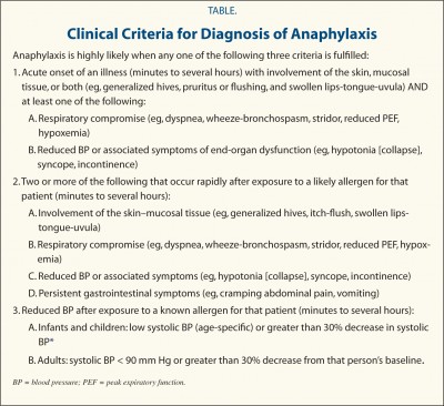
  • Anaphylactic shock is defined as blood pressure 30% below the patient’s baseline or a systolic blood pressure below 90 mm Hg. The systolic blood pressure is the top number when you get your blood pressure checked. If that top number is below 90 mm Hg, and that is the result of anaphylaxis, you are in anaphylactic shock.
  • Anaphylactic shock is the most serious potential complication of anaphylaxis. Anaphylactic shock happens when the chemicals released by mast cells cause a lot of the fluid in the bloodstream to “fall out” of the bloodstream and get stuck in the tissues.
  • When this happens, that fluid loss causes the blood pressure to drop. In response, the heart beats faster to try and use the blood it still has left to get oxygen to the body. However, at a certain point, even beating really fast is not enough to get enough blood to the tissues. At this point, shock sets in.
  • Anaphylactic shock occurs specifically as a result of low blood pressure. Because of this, providers strongly associate low blood pressure with anaphylaxis. They may not realize that while a person with high blood pressure cannot be having anaphylactic shock, they can be having anaphylaxis.
  • Part of the confusion is that anaphylaxis has been defined lots of different ways by many different groups. I have written a very detailed post about this (see the link below). Even today, exactly what constitutes anaphylaxis not agreed upon by everybody.
  • The most widely used criteria in the US are the criteria published in 2006 by the World Allergy Organization journal. These criteria explicitly state that a person does not need to have low blood pressure to be having anaphylaxis. A person can meet these criteria based upon a variety of combinations of symptom and vital signs that do not include low blood pressure.
2006 WAO Anaphylaxis Criteria

For additional information, please visit the following posts:

The definition of anaphylaxis
Anaphylaxis and mast cell reactions

The MastAttack 107: The Layperson’s Guide to Understanding Mast Cell Diseases, Part 49


The MastAttack 107: The Layperson’s Guide to Understanding Mast Cell Diseases, Part 62

76. Is it true that allergic reactions can cause heart attacks?

  • Yes.
  • Kounis Syndrome is an acute coronary syndrome caused by activated mast cells releasing chemicals. It is sometimes referred to as “allergic heart attack.” In acute coronary syndrome, there is not enough blood being pumped into the heart. It is named for two of the large blood vessels supplying oxygen to the heart, the coronary arteries. When not enough blood is getting to the heart via the coronary arteries, it can damage heart muscle, sometimes permanently. Heart attack and angina are examples of acute coronary syndromes.
  • In Kounis Syndrome, mast cells become activated, releasing lots of chemicals. These chemicals can irritate the coronary artery, causing it to spasm. This spasm reduces the amount of blood getting to the heart. Sometimes, mast cell activation can trigger the formation of a clot. A clot can be the reason not enough blood is passing through the artery.
  • Several of the molecules released by mast cells can affect the cardiovascular system and contribute to causing Kounis Syndrome. Histamine and leukotrienes can cause the coronary artery to narrow. It can also activate platelets, helping a clot to form. Both tryptase and chymase can cause clots formed elsewhere to break off and get stuck in the coronary artery.
  • Mast cells also help regulate an important molecule called angiotensin II. Angiotensin II is a powerful regulator of blood pressure and can cause the coronary artery to narrow and tighten up.
  • People with Kounis Syndrome may have a history of coronary artery disease. Some patients have a stent in the coronary artery from a previous coronary issue. A stent is a tube that helps keep the blood vessel the right size so that the heart gets the blood it needs. However, many patients with Kounis Syndrome do not have any history of problems with their heart or blood vessels.
  • The symptoms of Kounis Syndrome sometimes look just like the symptoms of any other mast cell reaction or anaphylaxis, making it hard to know that a person is having Kounis Syndrome. Chest pain, irregular heart beat, the heart beating too fast or too slow, and palpitations are all common symptoms of Kounis Syndrome.
  • Another tricky thing about Kounis Syndrome is that it doesn’t always show up on the tests we use to look for heart attack or coronary issues. Because of this, doctors don’t always realize what is happening. Some people do have positive results to these tests, things like EKG, echocardiogram, chest x-ray, and bloodwork to look at levels at cardiac enzymes or troponin. Cardiac enzymes and troponins are often high in a person who is having a heart attack but are sometimes normal for patients with Kounis Syndrome.
  • In order to manage Kounis Syndrome, patients may need treatment for both the allergic reaction and the coronary syndrome.
  • Treatment for the allergic reaction is similar to anaphylaxis treatment: an H1 antihistamine like Benadryl, an H2 antihistamine like famotidine, a corticosteroid like methylprednisolone, IV fluids, and sometimes epinephrine, if that’s appropriate. Please note that epinephrine is not always appropriate for patients who have Kounis Syndrome because epinephrine can actually also cause the coronary artery to narrow.
  • Treatment for the cardiovascular aspect of Kounis Syndrome is very dependent upon symptoms and test results. Calcium channel blockers like verapamil, aspirin, and nitroglycerin are commonly used. Importantly, some of the common medications used to manage coronary syndrome are not safe for mast cell patients. These medications include beta blockers like metoprolol or atenolol, and, to a lesser extent, ACE inhibitors like lisinophil. These medications can interfere with epinephrine so epinephrine may not work if a patient needs it for anaphylaxis.
  • Anything that triggers mast cell activation can cause Kounis Syndrome, including emotional stress.

For additional information, please visit the following posts:
Kounis Syndrome: Subtypes and effects of mast cell mediators (Part 1 of 4)
Kounis Syndrome: Diagnosis (Part 2 of 4)
Kounis Syndrome: Treatment (Part 3 of 4)
Kounis Syndrome: Stress (Part 4 of 4)
Beta blockers and epinephrine
Cardiovascular manifestations of mast cell disease: Part 1 of 5
Cardiovascular manifestations of mast cell disease: Part 2 of 5
Cardiovascular manifestations of mast cell disease: Part 3 of 5
Cardiovascular manifestations of mast cell disease: Part 4 of 5
Cardiovascular manifestations of mast cell disease: Part 5 of 5
The Provider Primers Series: Medications that impact mast cell degranulation and anaphylaxis