Skip to content

mastocytosis

The MastAttack 107: The Layperson’s Guide to Understanding Mast Cell Diseases, Part 81

94. How are mast cells involved in cancer?

  • Mast cells are very involved in cancer biology. They are frequently found in tumors. Tumors can trick mast cells into doing things they need to stay alive, like make blood vessels to supply the tumor with blood, and tissue remodeling, to push aside the healthy tissue and make room for the tumor.
  • Cancer is mast cell activating. All cancers. This is because cancers often trick the body into doing things that help the cancer and not the body, like I just described above. Having cancer frequently causes allergy symptoms because of mast cell activation.
  • Cancer can also cause the body to make more mast cells than normal, a condition called mast cell hyperplasia. This can happen because the body is trying to fight off the cancer with more immune cells or because it has been tricked by the cancer to make more mast cells to help the cancer.
  • Please note that mast cell hyperplasia is NOT the same as mastocytosis. Mast cell hyperplasia is too many healthy mast cells that function normally. Mastocytosis is too many aberrant mast cells that do not function normally. Cancer does not cause mastocytosis.
  • Long term inflammation increases future risk of cancer at the site of inflammation. This applies almost universally. Mast cells participate significantly in inflammation so they can contribute to the risk of cancer. For example, patients with long term colon inflammation, which may be caused by mast cells, are at increased risk of colon cancer.
  • Patients with mastocytosis have increased risk of developing cancer, especially those with systemic mastocytosis. As many as 40% of patients with systemic mastocytosis develop another blood disorder with too many broken cells. Frequently, the other blood disorder is a blood cancer like chronic myelogenous leukemia.
  • It is not yet known if mast cell activation carries an increased risk of developing cancer.
  • Two forms of systemic mastocytosis are cancerous, mast cell leukemia and mast cell sarcoma. These are both extremely rare and it is extremely rare for a person with a history of mast cell disease to develop either of these conditions.

For further reading, please visit the following post:

The MastAttack 107: The Layperson’s Guide to Understanding Mast Cell Diseases, Part 48

Mast cells in the GI tract: How many is too many? (Part Three)
The MastAttack 107: The Layperson’s Guide to Understanding Mast Cell Diseases, Part 59

73. Can mast cell disease cause organ damage?

  • Yes.
  • The term organ damage is tricky because people use it to mean a lot of things while providers and researchers often use it to mean one very specific thing. For providers and researchers, the term “organ damage” usually means a change in the organ that affects its structure, like it becomes misshapen or deformed in some way. Structural changes like this are often irreversible. This damage to the organ’s shape and structure usually affects how the organ works, called organ function.
  • When patients and laypeople talk about organ damage, they usually mean a change in the way the organ functions, even if the structure is not changed at all. This is different in a very important way: changes in an organ that do not affect its permanent structure can sometimes be reversible.
  • Both cutaneous and systemic mastocytosis cause organ damage in a way that damages the organ’s structure. When too many mast cells burrow into the tissue of an organ, it has to push other things out of the way. When you have mastocytosis, the mast cells like to stick together and form a big clump in the tissue. This punches holes in the tissue, affecting the organ’s structure and shape. This is called dense infiltration. It is one of the criteria for systemic mastocytosis and also happens in cutaneous mastocytosis.
  • In patients with mastocytosis, those mast cells clumping together cause a lot of the organ damage. This means that people who have the most mast cells usually have the worst organ damage. Patients with malignant forms of mast cell disease, like mast cell leukemia or aggressive systemic mastocytosis, often have organs that are riddled with TONS of mast cells.
  • Mast cells don’t live in the blood so when your body makes way too many mast cells, those mast cells will dive into whatever organ they can to get out of the bloodstream. This causes damage to the structure that you can see with scans or in biopsies.  People with mast cell leukemia and aggressive systemic mastocytosis suffer so much damage to the shape and function of their organs that the organs can totally stop working, called organ failure.
  • One of the key differences researchers and providers see between mastocytosis and mast cell activation syndrome is that mast cells don’t cause THIS TYPE of structural damage in mast cell activation syndrome patients.
  • We know this because in biopsies, they do not have mast cells clumped together to punch holes in the tissue. Sometimes they have lots of mast cells, but it is much less damaging to the tissue if they aren’t clumped together. Think of it like poking something with finger versus punching with your fist.
  • In MCAS, mast cells do not cause structural damage to organs IN THIS WAY. However, many people with MCAS do have structural damage to their organs. Many of them also have organs that do not function correctly even if the organs look normal.
  • Even if you don’t have mast cells punching holes in all your organs, they can still do a lot of damage. This is because mast cells cause lots of inflammation, which can stress out your organs. Over time, your organs can be damaged by the mast cells releasing too many mediators. While this is not always dangerous, it is certainly painful and frustrating.
  • Many MCAS and mastocytosis patients have a lot of damage to their GI tracts from years of vomiting, obstructions, diarrhea or constipation. Hives and mastocytosis spots can damage your skin, causing discoloration, scarring or sensitivity. Muscles can become weaker over time because of mast cell inflammation. Swelling can stretch out your skin and connective tissues. Nerves can be damaged significantly, affecting organ function. Bones can become brittle and break, or can become too dense because the body is making new bone when it shouldn’t.
  • All of these effects on organ function can be caused by mast cells. Major changes in organ function can also cause secondary conditions to arise.
  • Mast cell patients are also at an increased risk for anaphylaxis which can cause changes in organ function or organ damage.
  • Patients who have trouble breathing or low blood pressure may not be getting enough oxygen to their whole body. That can cause lasting damage if it goes on long enough.

For more detailed reading, please visit the following posts:

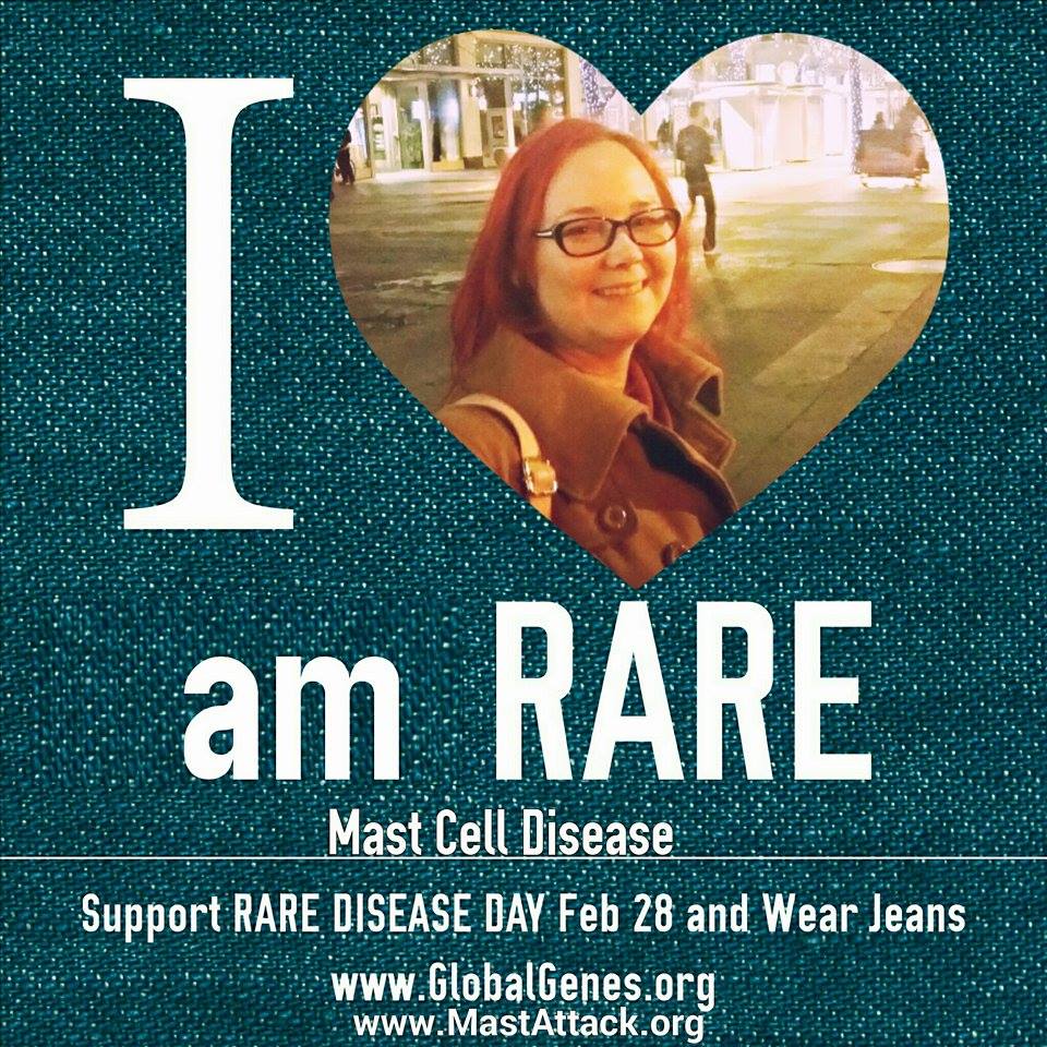
I am rare

A year ago this week, I started writing regular posts about mast cell disease and chronic illness. In honor of Rare Disease Day, the last day of February, I decided to put up short posts on Facebook daily for the remaining days of February. I could not have predicted that this would eventually give way to a website that is visited thousands of times a month by people all over the world.

I wanted to write a post about having a rare disease and what it meant to be a rare patient, but I have actually been too busy dealing with my rare disease to do it. This week, it occurred to me that I actually have multiple rare diseases. Today, I learned that four of my diagnoses are classified as rare diseases in the US. I have four individual rare diseases. This is not uncommon for mast cell patients.

In the US, any disease that affects less than 200,000 at one time is considered rare. These diseases can be infectious diseases, cancers, genetic disorders, autoimmune diseases, and so on. Rare diseases are defined differently by different countries and organizations. Likewise, a disease can be rare in one region and common in another.

There are over 7000 known rare diseases. Worldwide, they affect 300,000,000 people. In the US, they affect 25,000,000. If all rare diseases live together in one country, it would be the third most populous country in the world.

Almost 10% of the American population has at least one rare disease. 2/3 of Americans living with rare disease are children. Currently, only 350 rare diseases have an FDA approved treatment. This means that most of the medications we use are not designed for us and we don’t know how they will affect us.

Almost half of primary care physicians in the US say they feel uncomfortable with taking on a rare disease patient. It can take us up to six years to receive a correct diagnosis. Some people are never diagnosed.

80% of rare disease patients have one of 350 rare diseases, with the rest being significantly more rare. Mastocytosis is not one of those 350 diseases.


My name is Lisa Klimas. I am 31 years old and I live with four rare diseases.

Mast cell disease causes severe allergic reactions to things I am not actually allergic to.

Ehlers Danlos Syndrome causes hernias, joint instability, and poor wound healing.

Postural Orthostatic Tachycardia Syndrome causes dysregulation of blood pressure and heart rate.

Mixed connective tissue disease causes autoimmune activity against various tissues in my body.

All of these conditions are chronic, incurable, and painful.  Together they can cause life threatening complications.

February 28th is Rare Disease Day. For many people, it is just another day. But for me, it is a celebration.

It is a reminder that there are other people like me all over the world.

Alone, we are rare, but together we are many.  We are strong.  We are an army.

My name is Lisa Klimas and I am rare.


I am rare


*All figures from the National Organization for Rare Disorders (NORD).

MCAD: General information for public

Mast Cell Activation Disorders (MCAD): Frequently Asked Questions

What are mast cell activation disorders?

They are a group of conditions  in which the mast cells in the body do not function correctly.  MCAD includes systemic mastocytosis, urticaria pigmentosa and mast cell activation syndrome, among other conditions. Mast cells are responsible for allergic responses. In MCAD, patients can have allergic type reactions to things they are not allergic to. These reactions can be very severe and even life threatening.

What are mast cell reactions?

These are reactions caused by mast cells being improperly activated. These reactions vary from person to person. Symptoms can include, but are not limited to, nausea, vomiting, hives, rashes, itching, flushing (turning red), dizziness, confusion and irritability. Symptoms are caused by the chemicals released by the mast cells.

What causes mast cell reactions?

Triggers vary from person to person. More common triggers include heat, cold, friction (especially on the skin), sunlight, foodstuffs, physical exertion, stress, dyes and fragrances. Triggers can also change over time, with new triggers presenting.

Are mast cell reactions dangerous?

YES. Many MCAD patients will experience uncomfortable reactions throughout their lives. However, every reaction carries the risk of anaphylaxis, a life threatening, severe allergic reaction. Therefore, avoiding reactions as much as possible is very important for mast cell patients. Each patient has an individualized response plan. For many, it involves removal of trigger and administration of medication, such as antihistamines or inhalers.

What is anaphylaxis?

Anaphylaxis is a severe allergic reaction affecting multiple organ systems in the body. These are the kinds of reactions observed in patients with bee sting allergies. Anaphylaxis can be fatal. It is a medical emergency requiring immediate treatment, usually epinephrine (Epipen.) Please receive guidance from treating physician on when to use an Epipen.

How are mast cell anaphylaxis and mast cell reactions different from normal allergies (like food allergies?)

With allergies, your body reacts by a specific method that involves ingesting and recognizing the allergen. In MCAD patients, the mast cells incorrectly think many things are allergens. Since mast cells are so sensitive in these people, ingestion of an allergen is NOT necessary to cause mast cell reactions or anaphylaxis. Smelling a perfume or breathing in very hot, humid air is enough to cause a reaction in many MCAD patients.

What causes MCAD?

Genetic mutations cause different kinds of MCAD. Recent studies have shown that mast cell disease can affect multiple members of the same family.

Why do some MCAD patients have spots?

These spots occur in locations where there are more mast cells than usual in the skin. These are NOT contagious rashes. In addition, MCAD patients who do not have permanent spots often have very sensitive skin, which may cause temporary marks or rashes.

How can I help an MCAD patient be safe?

By not being afraid of their disease. Respect their triggers and help them work around these limitations. Reactions can be painful and very scary, especially for kids. Learn the symptoms associated with reactions and be ready to help with a response plan.


Is there more information you feel should be included here?  Let me know in the comments and I can add it in.

Pregnancy in mastocytosis

Mast cells are involved in regulating the female reproductive cycle.  There are direct correlations between the contraction of uterine wall and serum histamine during pregnancy, as well as correlations to other mast cell mediators.  Mast cell activation and degranulation in the endometrium occurs immediately before and during menses.  Many allergic conditions, including asthma and angioedema, are exascerbated during menses, which is thought to be due to mast cell activation.

Throughout pregnancy, sex hormone levels change.  Sex hormones, such as estrogen, can directly influence mast cell activation and degranulation.  Mastocytosis patients often discontinue antihistamine and antimediator medications during pregnancy due to safety concerns.  In 2011, a study was published examining the effect of pregnancy on mastocytosis patients and of mastocytosis on pregnancy and delivery.
During pregnancy, 45% had itching; 40% had flushing; 24% had GI symptoms; and 9% had anaphylaxis.  22% of patients reported worsening symptoms throughout the pregnancy.  2% felt their symptoms were more frequent, while 18% developed new symptoms, and 2% had both new and more frequent symptoms.  New symptoms generally appeared in the first trimester, occasionally in the third.  Worsening of symptoms occurred in 3/6 women with CM and 7/35 with ISM with skin involvement.  One woman developed skin lesions during the third trimester and was diagnosed with ISM via bone marrow biopsy after delivery.
33% of women reported their symptoms had improved during pregnancy.  15% had complete resolution of symptoms, 15% had at least one symptom disappear and 3% had at least one symptom disappear but others worsen.  All resolutions occurred during the first trimester and lasted throughout the pregnancy with the exception of one patient.  In patients who had idiopathic anaphylaxis before pregnancy, 50% of them had no anaphylaxis while pregnant.  In those women who did have anaphylaxis during pregnancy, it was resolved without the use of epinephrine and did not cause early labor or complications.
Complete resolution of symptoms occurred in a patient with well differentiated SM (WDSM), ½ patients with ISM and no skin involvement, 9% of patients with ISM with skin involvement and 17% of patients with CM.  Partial resolution occurred in ½ patients with ISM and no skin involvement, 11% of patients with ISM with skin involvement and 17% of patients with CM.  In 6% of patients with ISM with skin involvement, at least one symptom disappeared while others worsened. 
45% had no change in symptoms during pregnancy (19/35 ISM with skin involvement patients, 1/6 CM patients.)  One patient experienced significant improvement of skin lesions in the first trimester. 
For women who worsened during pregnancy, mast cell symptoms continued to be worse after delivery for 50% of them.  Symptom resolution observed during pregnancy continued after delivery for 4/7 cases that had complete resolution and 3/6 cases that had partial resolution.  Complete resolution of symptoms occurred in two patients that had partial resolution during pregnancy.  There were five cases of worsening skin lesions after delivery. 
78% of infants were delivered vaginally and 22% by Caesarean.  Nine deliveries were induced with oxytocin (8/9) or dinoprostone (1).  In 38% of cases, the patient took mast cell pre-meds at the onset of labor.  Anesthesia was used in 82% of cases, including epidural (32 cases), local (3) and general (2.)  11% of patients had mast cell attacks during or immediately after labor.  Anesthesia and medications used for labor seemed to be safe.  Only three women had mast cell reactions to epidural; of these, two had not take pre-meds.  Premedication at the initiation of labor is recommended.
3 out of 45 newborns were born premature and 4 out of 45 had low birth weights.  One had Down Syndrome; one had respiratory distress; one had jaundice; and one had heart rhythm abnormalities before birth.  There was no correlation between mother’s symptoms and outcome.  There was no correlation between anaphylaxis and outcome.  None of the children had skin lesions at birth, but one developed CM at the age of 5. 
The frequency of spontaneous pregnancy loss during first trimester (10-15), birth by Caesarean section (25%), prematurity (7.6%) and low birth weight (3-5.8%) were comparable to the rates in the general population.  Nonaggressive forms of mastocytosis did not appear to impact pregnancy outcome.
Reference:
Matito, A., et al.  Clinical Impact of Pregnancy in Mastocytosis: A Study of the Spanish Network on Mastocytosis (REMA) in 45 Cases.  Int Arch Allergy Immunol 2011;156:104-111.

On prognosis and dying from mast cell disease


There isn’t a lot of data on death from mast cell disease.  Not real data, with statistics and numbers.  People with SM and MCAS are frequently reassured that they will live a normal life span.  People with SM-AHNMD are quoted an average survival of about 8.5 years; ASM, 3.5 years; MCL, under a year. 
Of those groups, only the survival time for mast cell leukemia is convincing to me.  This is because mast cell leukemia has a pretty homogenous presentation, meaning that it affects most people in the same way.  When a disease is as rare as MCL, it is important that you remove as many variables as possible in order for the data to be sound.  And that’s the problem with the rest of the survival data, to my eyes – there’s just too much variability.  Throw in a patient population as small as ours and you’ve got a lot of uncertainty.
The effects of mast cell disease are highly individualized.  There are several B and C findings, meaning that combinations of symptoms and manifestations are very variable.  The SM-AHNMD group is a good example of this.  This category lumps together many different combinations of diseases, not to mention the stages of those diseases.  Someone with ASM-AML is going to have a very different prognosis than someone with SM-CEL.  Simply averaging the lifespans of these people and quoting this as a life expectancy does the mast cell community a disservice.  It is important to remember this when you are typing “mast cell disease death” in the middle of the night. 
Even though we know that most people with SM die from something else, or that for many people, it is a very manageable disease, there is always the possibility that it will be different for you.  It’s hard not to imagine that you will be in the unlucky percentage of people that have progressive disease, that develop ASM, that have leukemic transformation.  Admonishing people who bring up this concern as “negative” or “paranoid” doesn’t make it less terrifying.  It just makes people more afraid to talk about the fact that sometimes people die from mast cell disease and often they aren’t sure how best to minimize their chances of becoming one of them.
Due to the differences in presentation, it has been difficult to identify markers that definitively indicate prognosis.  A lot of effort was put into looking at various CKIT mutations, not just D816V, to see if this could be predictive.  There has not been statistically significant data that this is the case.
The closest things we have to prognostic markers don’t get a lot of play in the general mast cell consciousness.  We talk a lot about CKIT because it affects treatment, and symptoms because it affects diagnosis.  But beyond the initial workup, we don’t often hear much about the CD2 and CD25 markers.  However, a paper published in 2009, established a link between “immunophenotype,” in this case which markers the cells present, and prognosis. 
This study looked at bone marrow samples from 123 patients with different types of SM, including MCL.  Importantly, they also had a large control group of people who did not have SM.  A solid control group is key to determining that a finding is real.  They defined the patients as either good-prognosis (SM, well differentiated SM, and cMAD, clonal mast cell activation disorder (what we now call monoclonal mast cell activation syndrome, MMAS)), or poor-prognosis (ASM and MCL.) 
They determined that for patients whose mast cells expressed BOTH CD25 and CD2 (ISM/MMAS) or NEITHER CD25 and CD2 (WDSM), prognosis was good.  However, mixed expression (typically CD25+ and CD2-) indicated a poorer prognosis.  They compared it to current markers, like the D816V mutation and serum tryptase, as well as clinical findings, like swollen spleen, swollen liver, skin lesions and white blood cell count.  The expression of markers was found to be a sounder method for estimating life expectancy than any of these.
It’s okay to be scared.  We all know people who have died from mast cell disease.  It is scary to think that we could be next.  It is scary to live under the looming threat of anaphylaxis.  But the good news is that science is trying to catch up.  More people are being diagnosed with mast cell disease, and science is getting better at identifying the ways that we are alike and different.  There is every reason to think we will have comforting data in the future.  We just have to get there. 


Reference:
Teodosio, Cristina, et al.  2009.  Mast cells from different molecular and prognostic subtypes of systemic mastocytosis display distinct immunophenotypes.  Journal of Allergy and Clinical Immunology, 125: (3), 719-726.